Noninvasive Optical Imaging Technique Monitors Postprandial Cardiovascular Health
Posted on 26 Sep 2024
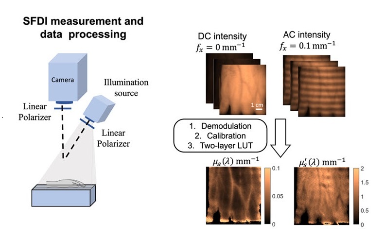
The dynamics of blood nutrient and lipid levels following a high-fat meal are key indicators of both current and future cardiovascular health. Traditionally, these levels have been measured through invasive blood draws, which are not practical for routine health monitoring. To address this, researchers are investigating noninvasive methods that could enhance the tracking of post-meal effects and help identify contributors to cardiovascular disease. One promising technique is a noncontact optical imaging method called "spatial frequency domain imaging" (SFDI), which measures tissue properties and blood flow dynamics.
In a new study, a research team that included scientists from Boston University (Boston, MA, USA) explored how different meal compositions affect skin tissue properties immediately after consumption. They focused on the peripheral tissue of the hand to observe the short-term effects of both low-fat and high-fat meals. The team used SFDI to monitor 15 subjects who consumed both types of meals on separate occasions. They imaged the back of each participant's hand every hour for five hours post-meal, analyzing three specific wavelengths to assess hemoglobin, water, and lipid concentrations. The findings revealed notable differences in tissue responses. After a high-fat meal, tissue oxygen saturation increased, while the low-fat meal caused a decrease, indicating that dietary fat not only impacts long-term health but also triggers immediate physiological changes. The peak changes occurred three hours post-meal, aligning with spikes in triglyceride levels, as reported in Biophotonics Discovery (BIOS).

In addition to imaging, the researchers monitored blood pressure and heart rate, and conducted blood draws to measure triglycerides, cholesterol, and glucose levels. The study showed that changes in optical absorption at specific wavelengths corresponded closely with variations in lipid concentrations. Using this data, the team developed a machine learning model to predict triglyceride levels from the SFDI measurements, achieving accuracy within 40 mg/dL. This advancement could pave the way for noninvasive monitoring of cardiovascular health.
“The research suggests that SFDI could serve as a promising alternative, allowing for easier monitoring of how meals affect cardiovascular health,” said senior author Darren Roblyer, professor of biomedical engineering at Boston University. “Overall, these findings highlight the intricate relationship between diet, body response, and cardiovascular risk, suggesting a need for further exploration of non-invasive assessment methods.”


