Orthopedic Surgeons Harness Power of 3D Printing
By HospiMedica International staff writers
Posted on 16 Aug 2017
A new study shows how patient-specific three-dimensional (3D) models can be used to overcome steep learning curves for complex surgical procedures.Posted on 16 Aug 2017
Researchers at the University of California, San Diego (UCSD; USA) and Rady Children's Hospital (San Diego; CA, USA) conducted a study in 15 children who underwent a three-plane proximal femoral osteotomy (TPFO) for symptomatic proximal femoral deformity due to a slipped capital femoral epiphysis (SCFE). Five of the patients underwent preplanned surgery using 3D-printed models; the other five underwent the same procedure with the same surgeon, but without the models. In addition, two other surgeons operated on a different group of five patients, without using models.
.")
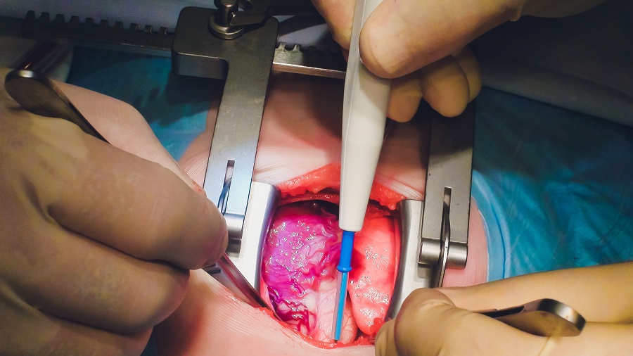
Image: Patient-specific 3D models aid in surgical planning of complex 3D orthopedic surgery (Photo courtesy of the UCSD).
The results showed that when using the 3D-printed models, surgical time decreased by 45 minutes and 38 minutes, and fluoroscopy time decreased by 50% and 25%, in the model group compared with the no-model and senior groups, respectively. According to the researchers, the time saved could translate into at least USD 2,700 in savings per surgery. By contrast, after the one-time cost of buying a 3D printer for about USD 2,200, physicians can make a model for each surgery for about USD 10. The study was published in the April 2017 issue of the Journal of Children's Orthopaedics.
“Being able to practice on these 3D-models is crucial,” said senior author pediatric orthopedic surgeon Vidyadhar Upasani, MD, of Rady Children's Hospital. “The results of the study were so positive that Rady Children's orthopedics department has acquired its own 3D printer. I've seen how beneficial 3D models are; it's now hard to plan surgeries without them.”
SCFE is a condition in which the head of the femur slips along the bone's growth plate, deforming it. The main goal of surgery is to sculpt the femur back into its normal shape and restore hip function. This is difficult because the bone and its growth plate are not directly visible, so that surgeons cannot visualize in 3D how the growth plate is deformed. Physicians therefore study X-rays of the surgery site to plan the bone cuts; during surgery, X-ray fluoroscopy is used for guidance. These methods are time consuming and expose the child to radiation. In addition, physicians don't have a physical model to educate patients or practice the surgery beforehand.
Related Links:
University of California, San Diego
Rady Children's Hospital
Gold Member
STI Test
Vivalytic Sexually Transmitted Infection (STI) Array

Gold Member
12-Channel ECG
CM1200B

Silver Member
Wireless Mobile ECG Recorder
NR-1207-3/NR-1207-E
New
LED Phototherapy System
Bililed Mini+