


Spectral Domain OCT System Incorporates High Resolution Fundus Camera
|
By HospiMedica International staff writers Posted on 18 Jan 2010 |
|
The fundus, or inner lining, of the eye includes the retina and the blood supply to it. It can be photographed with specially designed cameras through the dilated pupil of the patient.
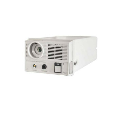
The 3D OCT-2000 system is an optical signal acquisition and processing method. A spectral domain optical coherence tomography (OCT) system incorporates a high-resolution fundus camera and a user-friendly color touch-screen display.
A product of Topcon (Tokyo, Japan), the new 3D OCT-2000 system has a compact, space-saving design. It captures micrometer-resolution, three-dimensional (3D) images from within optical scattering media (e.g., biological tissue). Commercially available OCT systems are employed in diverse applications, such as in diagnostic medicine, notably in ophthalmology, where it is used to obtain detailed images from within the retina.
Intuitive FastMap software enables dynamic viewing of the OCT data and provides 3D, 2D, and fundus images simultaneously. Pinpoint registration indicates the location of the OCT image within the fundus image. In addition, the compare function allows users to view serial exams in a comparison view and apply different analytical tools.
The seamless integration of 3D OCT-2000 with Topcon's Eye Route Image Management System provides connectivity and access to images anywhere, at anytime. "At Topcon we strive to be the leading provider of clinical information, technology and connectivity. The multi-touch experience that we are pioneering, underlines our visionary approach in enhancing workflow and communication within the ophthalmic industry,” commented Katrin Teigeler, vice president, marketing at Topcon.
Topcon Medical Systems, Inc. (TMS) is a supplier of ophthalmic diagnostic equipment and image management solutions.
Related Links:
Topcon
Gold Member
STI Test
Vivalytic Sexually Transmitted Infection (STI) Array
New
Gas Analyzer
GE SAM
New
Wound Irrigation Solution
Prontosan®
Latest Surgical Techniques News
- Ultrasound Technology Aims to Replace Invasive BPH Procedures
- Continuous Monitoring with Wearables Enhances Postoperative Patient Safety
- New Approach Enables Customized Muscle Tissue Without Biomaterial Scaffolds
- Robot-Assisted Brain Angiography Improves Procedural Outcomes
- Brain Mapping Technology Enhances Precision in Brain Tumor Resection
- Handheld Robotic System Expands Options for Total Knee Surgery
- VR Experience Reduces Patient Anxiety Before Kidney Stone Procedure
- Injectable Mini Livers Offer Hope for Patients Awaiting Transplant
- Pulsed Field Ablation Technology Cleared in Europe for Persistent AFib
- AI-Powered Imaging Brings Real-Time Margin Clarity to Breast Cancer Surgery
- Minimally Invasive Device Safely Treats Challenging Brain Aneurysms
- Surgical Robot Makes Complex Liver Tumor Surgery Safer and Less Invasive
- Neurostimulation Implant Reduces Seizure Burden in Drug-Resistant Epilepsy
- Minimally Invasive Procedure Effectively Treats Small Kidney Cancers
- Fluorescence Probe Paired with Engineered Enzymes Lights Up Tumors for Easier Surgical Removal
- Novel Hydrogel Could Become Bone Implant of the Future
Channels
Artificial Intelligence
view channelAI Analysis of Pericardial Fat Refines Long-Term Heart Disease Risk
Accurately identifying long-term cardiovascular disease risk in asymptomatic adults remains challenging for clinicians. Missed or underestimated risk delays preventive therapy and increases the chance... Read more")
Machine Learning Approach Enhances Liver Cancer Risk Stratification
Hepatocellular carcinoma, the most common form of primary liver cancer, is often detected late despite targeted surveillance programs. Current screening guidelines emphasize patients with known cirrhosis,... Read more")
, k-means++ was used to identify clusters. Three clinically distinct clusters were identified. Among those clusters, patients were divided into solid organ transplant (SOT) and non-SOT groups. Clinical characteristics and outcomes were evaluated (Masayuki Nigo et al., American Journal of Transplantation (2026). DOI: 10.1016/j.ajt.2025.10.019)")
Critical Care
view channel")
Noninvasive Monitoring Device Enables Earlier Intervention in Heart Failure
Hospitalizations for heart failure with preserved ejection fraction (HFpEF) remain common because lung congestion often worsens before symptoms prompt treatment changes. Missed early decompensation... Read more")
Automated IV Labeling Solution Improves Infusion Safety and Efficiency
Medication administration in high-acuity settings is often complicated by multiple concurrent infusions, making accurate line identification essential. In a 10-hospital intensive care unit study, 60% of... Read more)")
")
Surgical Techniques
view channel")
Ultrasound Technology Aims to Replace Invasive BPH Procedures
Benign prostatic hyperplasia (BPH) is a frequent cause of lower urinary tract symptoms in aging men and often requires invasive procedures or prolonged recovery. With prevalence expected to rise as populations... Read more")
Continuous Monitoring with Wearables Enhances Postoperative Patient Safety
Postoperative hypoxemia on general surgical wards is common and often missed by intermittent vital sign checks. Undetected low oxygen levels can delay recovery and raise the risk of complications that... Read more")
")
Patient Care
view channel")
Wearable Sleep Data Predict Adherence to Pulmonary Rehabilitation
Chronic obstructive pulmonary disease (COPD) is a long-term lung disorder that makes breathing difficult and often disturbs sleep, reducing energy for daily activities. Limited engagement in pulmonary... Read more")
Revolutionary Automatic IV-Line Flushing Device to Enhance Infusion Care
More than 80% of in-hospital patients receive intravenous (IV) therapy. Every dose of IV medicine delivered in a small volume (<250 mL) infusion bag should be followed by subsequent flushing to ensure... Read more")
")
Health IT
view channel")
EMR-Based Tool Predicts Graft Failure After Kidney Transplant
Kidney transplantation offers patients with end-stage kidney disease longer survival and better quality of life than dialysis, yet graft failure remains a major challenge. Although a successful transplant... Read more")
Printable Molecule-Selective Nanoparticles Enable Mass Production of Wearable Biosensors
The future of medicine is likely to focus on the personalization of healthcare—understanding exactly what an individual requires and delivering the appropriate combination of nutrients, metabolites, and... Read more")
")
")
Business
view channel")
")
")