


Virtual Autopsy Table Extends Anatomical Education
|
By HospiMedica International staff writers Posted on 12 Dec 2012 |
|
.")
Image: The Anatomage virtual dissection table (Photo courtesy of Anatomage).
A virtual autopsy table provides life size visualization of full body anatomy, which could help change the way anatomy is taught in medical schools.
The Anatomage virtual dissection table delivers a realistic virtual cadaver with accurate anatomic details. The form factor of the Table allows students to replicate a true operating table experience, using realistic interactive visualization of human three-dimensional (3D) anatomies. The virtual patient is recumbent as students stand beside the table and interact with it, rotating or completely changing the view with the swipe of a finger. Students can also cut the body, peel off soft tissue, or remove an organ with finger gestures, and unlike cadavers, the students can redo and undo the dissection again and again.
The Table also allows students to visualize skeletal tissues, muscles, organs, and soft tissue. These various tissues and views can be customized by virtually slicing, layering, and segmenting the anatomy, adding a new dimension of depth to the education that students receive. Custom annotations can be easily added by the students to the visualizations of anatomical structures. With the flexible annotation tools, medical schools and institutions can create innovative programs, quizzes, and new methods of study.
The Table comes with a full body gross anatomy model rendered from computerized tomography (CT) scan data that is fused with anatomically accurate, textured surface models for educational purposes. The Table can also open any data from any CT, magnetic resonance imaging (MRI), and ultrasound scanners. Additional photographic images, or even full presentations, can also be displayed on the Table. In addition, by incorporating physical instruments into a curriculum, the Table provides a medium for simulating operation procedures in a natural and intuitive manner.
The Table hardware is based on a liquid crystal display (LCD) high-contrast screen with a 3,960 x 1,080 resolution. Optical touch interactive sensors are used for image manipulation, which can be used with a finger or a stylus. Around a dozen people can easily stand around and comfortably interact with each other and the Table at the same time. If needed, it can be moved to a different room on rollers that can be locked into a position. The Anatomage virtual dissection table is a product of Anatomage (San Jose, CA, USA).
Related Links:
Anatomage
Gold Member
SARS‑CoV‑2/Flu A/Flu B/RSV Sample-To-Answer Test
SARS‑CoV‑2/Flu A/Flu B/RSV Cartridge (CE-IVD)
Gold Member
Neonatal Heel Incision Device
Tenderfoot
Glucose Meter
StatStrip®
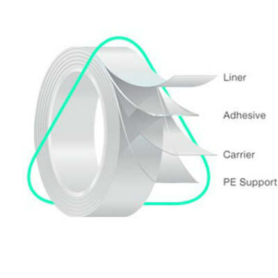
Medical Adhesive
MED 5570U
Latest Health IT News
- AI-Native EHR Achieves EU Medical Device Certification
- EHR-Integrated Screening Workflow Detects Cognitive Impairment at Admission
- AI System Detects and Quantifies Chronic Subdural Hematoma
- Continuous Monitoring Platform Detects Infection Risk Across Care Transitions
- Automated System Classifies and Tracks Cardiogenic Shock Across Hospital Settings
- Voice-Driven AI System Enables Structured GI Procedure Documentation
- EMR-Based Tool Predicts Graft Failure After Kidney Transplant
Channels
Artificial Intelligence
view channel")
AI Platform Supports Noninvasive Remote Hemodynamic Monitoring in Heart Failure
Heart failure remains a leading cause of hospitalization in adults over 65, affecting more than 6.7 million people in the U.S. Clinicians often lose visibility into hemodynamic deterioration once patients... Read more")
AI Tool Predicts Unplanned Care and Symptom Burden in Cancer Survivors
Unplanned emergency visits and hospitalizations remain common in cancer survivorship, when routine clinical contact often tapers while new symptoms emerge. These events reflect unmet needs and disrupt... Read more")
")
Critical Care
view channel")
Handheld ECG Algorithm Shows Promise for At-Home Heart Attack Risk Assessment
Chest pain remains one of the most common emergency presentations, yet determining which patients are experiencing a heart attack outside the hospital is challenging. Delays from symptom onset to hospital... Read more")
Smartphone Heart Rhythm App Reduces Unnecessary Cardioversion Procedures
Atrial fibrillation, an irregular and often rapid heart rhythm, is the most common arrhythmia in adults. Elective electrical cardioversion is frequently canceled on the day of treatment when patients revert... Read more")
")
Surgical Techniques
view channel")
New CAR T-Cell Therapy Enables Transplants in Hard-to-Match Kidney Patients
Highly sensitized kidney transplant candidates have extremely high levels of anti-donor antibodies that block matching and prolong dialysis. These patients face long wait times and increased morbidity... Read more")
CE-Marked Ultrasonic Shears Streamline Breast and Thyroid Surgery
Thyroid and breast surgeries are often performed in confined anatomical spaces near critical structures, making precise dissection and controlled thermal management essential. As the global disease burden... Read more")

Patient Care
view channel")
AI Avatar Doctor Improves Patient Understanding Before Radiotherapy
Radiation oncology consultations require patients to grasp complex concepts quickly, yet anxiety and information overload often undermine understanding and informed consent. Poor comprehension can also... Read more")
Wearable Sleep Data Predict Adherence to Pulmonary Rehabilitation
Chronic obstructive pulmonary disease (COPD) is a long-term lung disorder that makes breathing difficult and often disturbs sleep, reducing energy for daily activities. Limited engagement in pulmonary... Read more")
")
Point of Care
view channel")
Portable MRI System Accelerates Emergency Brain Imaging and Triage
Emergency departments frequently face delays accessing conventional magnetic resonance imaging (MRI) for patients with suspected neurological emergencies. Such waits can slow triage, prolong boarding,... Read more platform for obstetrics. (Photo courtesy of HemoSonics)")
")
")
")
")