


Pathology Device Averts Repeated Breast Cancer Surgeries
|
By HospiMedica International staff writers Posted on 16 Jul 2013 |
|
.")
Image: The prototype device applies an adhesive film to the breast tissue before examination (Photo courtesy of Will Kirk/homewoodphoto.jhu.edu).
A new tool will allow pathologists to quickly inspect excised breast tissue while the patient is still in the operating room (OR), reducing the need for a second surgery.
Developed by graduate students at Johns Hopkins University (Baltimore, MD, USA), the prototype device applies an adhesive film to the specimen breast tissue before it is sliced; the film thus holds the delicate tissue together, preventing damage to the samples during the preparation process. The result is a sample that can be clearly reviewed by a pathologist within 20 minutes of its removal, potentially eliminating the need for a second operation on another day. The low-cost system includes a reusable applicator and a proprietary disposable film.
“We spoke to breast cancer surgeons,” said Hector Neira, MSc, one of the student inventors. “They told us that they are desperate for something that will allow them to remove the tumor in its entirety the first time, so that the patient doesn’t have to come back for a second surgery.”
“I think the students have been incredibly creative in their development of this concept, and they are addressing a very real need in the field of breast cancer surgery,” added professor of Surgery Melissa Camp, who worked with the students. “Accurate assessment of margin status during the initial operation will lead to fewer reoperations, and this will be beneficial for patients in many respects. I look forward to their continued work!”
When most tumors are removed, pathologist can quickly flash-freeze the tissue and slice off paper-thin samples for microscopic examination; if the pathologist sees that cancer cells extend to the outer edge or margin of a sample, the surgeon is advised to remove more tissue from the patient. Breast tissue, however, poses a problem; it has a high fat content and does not freeze well, causing the samples to smear, form gaps, and become unsuitable for a quick review.
Related Links:
Johns Hopkins University
Gold Member
STI Test
Vivalytic Sexually Transmitted Infection (STI) Array
New
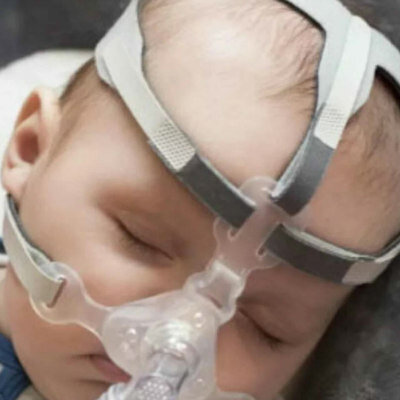
Pediatric Mask
Respire SOFT
New
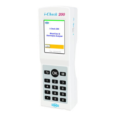
Blood Gas Analyzer
i-Check200
Latest Surgical Techniques News
- Ultrasound Technology Aims to Replace Invasive BPH Procedures
- Continuous Monitoring with Wearables Enhances Postoperative Patient Safety
- New Approach Enables Customized Muscle Tissue Without Biomaterial Scaffolds
- Robot-Assisted Brain Angiography Improves Procedural Outcomes
- Brain Mapping Technology Enhances Precision in Brain Tumor Resection
- Handheld Robotic System Expands Options for Total Knee Surgery
- VR Experience Reduces Patient Anxiety Before Kidney Stone Procedure
- Injectable Mini Livers Offer Hope for Patients Awaiting Transplant
- Pulsed Field Ablation Technology Cleared in Europe for Persistent AFib
- AI-Powered Imaging Brings Real-Time Margin Clarity to Breast Cancer Surgery
- Minimally Invasive Device Safely Treats Challenging Brain Aneurysms
- Surgical Robot Makes Complex Liver Tumor Surgery Safer and Less Invasive
- Neurostimulation Implant Reduces Seizure Burden in Drug-Resistant Epilepsy
- Minimally Invasive Procedure Effectively Treats Small Kidney Cancers
- Fluorescence Probe Paired with Engineered Enzymes Lights Up Tumors for Easier Surgical Removal
- Novel Hydrogel Could Become Bone Implant of the Future
Channels
Artificial Intelligence
view channelAI Analysis of Pericardial Fat Refines Long-Term Heart Disease Risk
Accurately identifying long-term cardiovascular disease risk in asymptomatic adults remains challenging for clinicians. Missed or underestimated risk delays preventive therapy and increases the chance... Read more")
Machine Learning Approach Enhances Liver Cancer Risk Stratification
Hepatocellular carcinoma, the most common form of primary liver cancer, is often detected late despite targeted surveillance programs. Current screening guidelines emphasize patients with known cirrhosis,... Read more")
, k-means++ was used to identify clusters. Three clinically distinct clusters were identified. Among those clusters, patients were divided into solid organ transplant (SOT) and non-SOT groups. Clinical characteristics and outcomes were evaluated (Masayuki Nigo et al., American Journal of Transplantation (2026). DOI: 10.1016/j.ajt.2025.10.019)")
Critical Care
view channel")
Noninvasive Monitoring Device Enables Earlier Intervention in Heart Failure
Hospitalizations for heart failure with preserved ejection fraction (HFpEF) remain common because lung congestion often worsens before symptoms prompt treatment changes. Missed early decompensation... Read more")
Automated IV Labeling Solution Improves Infusion Safety and Efficiency
Medication administration in high-acuity settings is often complicated by multiple concurrent infusions, making accurate line identification essential. In a 10-hospital intensive care unit study, 60% of... Read more)")
")
Patient Care
view channel")
Wearable Sleep Data Predict Adherence to Pulmonary Rehabilitation
Chronic obstructive pulmonary disease (COPD) is a long-term lung disorder that makes breathing difficult and often disturbs sleep, reducing energy for daily activities. Limited engagement in pulmonary... Read more")
Revolutionary Automatic IV-Line Flushing Device to Enhance Infusion Care
More than 80% of in-hospital patients receive intravenous (IV) therapy. Every dose of IV medicine delivered in a small volume (<250 mL) infusion bag should be followed by subsequent flushing to ensure... Read more")
")
Health IT
view channel")
EMR-Based Tool Predicts Graft Failure After Kidney Transplant
Kidney transplantation offers patients with end-stage kidney disease longer survival and better quality of life than dialysis, yet graft failure remains a major challenge. Although a successful transplant... Read more")
Printable Molecule-Selective Nanoparticles Enable Mass Production of Wearable Biosensors
The future of medicine is likely to focus on the personalization of healthcare—understanding exactly what an individual requires and delivering the appropriate combination of nutrients, metabolites, and... Read more")
")
")
Business
view channel")
")
")