Pioneering System Diagnoses Cancerous Tissue During Endoscopy
|
By HospiMedica International staff writers Posted on 16 Jun 2014 |
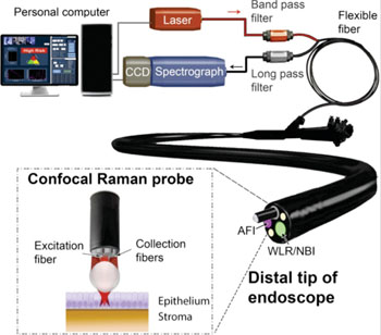
Image: Rapid fiber-optic confocal Raman spectroscopy system developed for real-time in vivo epithelial tissue diagnosis and characterization during endoscopy (Photo courtesy of Prof. Zhiwei Huang, the National University of Singapore, and the journal Gastroenterology).
A biomedical engineering team has developed a first of its kind in vivo molecular diagnostic system that makes highly objective, real-time cancer diagnosis during endoscopic examination a reality.
A National University of Singapore (NUS) team led by Associate Professor Huang Zhiwei, Department of Biomedical Engineering, has developed what is currently the only system clinically shown to be used in human patients for diagnosing even precancerous tissue in gastrointestinal tract during endoscopic examination in real time. Unlike conventional endoscopy that relies on the physician's visual interpretation of the images followed by a pathologist's analysis of the biopsy specimen several days later, their diagnostic system utilizes computer analysis of biomolecular information that can provide diagnosis in real time. It is a paradigm shift from a complex to a simple, objective, and rapid diagnostic procedure.
The In Vivo Molecular Diagnostic (IVMD) system is based on confocal Raman spectroscopy and includes a proprietary confocal fiber-optic probe connected to a customized online software control system. The fiber-optic probe enables the collection of biomolecular fingerprint of tissues in less than a second—while the online software enables this information to be extracted and analyzed, with diagnostic result presented during endoscopic examination. The IVMD system has been used in more than 500 patients in Singapore across diverse cancer types such as stomach, esophagus, colon, rectum, head and neck, and cervix. The researchers have also published more than 40 peer-reviewed publications, most recently a report by Bergholt MS, et al. in the journal Gastroenterology, published January 2014.
“We are delighted to not only overcome the technical challenges of weak Raman signal, high fiber background noise, and lack of depth perception by using our specially designed probe, but also to enable real-time diagnostic results to be displayed during endoscopy with our customized software,” said Prof. Huang.
For the clinical testing, the team has been collaborating with researchers from the NUS Yong Loo Lin School of Medicine, led by its Dean, Associate Professor Khay Guan Yeoh. Prof. Yeoh commented, “This remarkable new system is the first such diagnostic probe that can be used real-time, inside the human body, providing almost instantaneous information on cellular changes, including cancer and pre-cancer. This is a first in the world development, pioneered here in Singapore. It has the potential to make enormous clinical impact to how cancer is diagnosed and managed. The immediate point-of-care diagnosis during live endoscopic examinations will provide benefits in time and cost-savings, and will improve our patients’ prognosis.”
“It has been a long tedious journey of more than 10 years. The journey could be longer if not for the excellent cross-disciplinary teamwork at NUS. The contribution of the NUS clinical team is invaluable in demonstrating the clinical benefits of the system,” added Prof Huang. Moving forward, the team will conduct larger scale clinical trials, mainly in gastrointestinal cancer, to further validate the utility of this novel system.
Related Links:
National University of Singapore
Video: Clinical Use of Raman spectroscopy software during an endoscopic procedure
Latest Surgical Techniques News
- Neural Device Regrows Surrounding Skull After Brain Implantation
- Surgical Innovation Cuts Ovarian Cancer Risk by 80%
- New Imaging Combo Offers Hope for High-Risk Heart Patients
- New Classification System Brings Clarity to Brain Tumor Surgery Decisions
- Boengineered Tissue Offers New Hope for Secondary Lymphedema Treatment
- Dual-Energy Catheter Brings New Flexibility to AFib Ablation
- 3D Bioprinting Pushes Boundaries in Quest for Custom Livers
- New AI Approach to Improve Surgical Imaging
- First-Of-Its-Kind Probe Monitors Fetal Health in Utero During Surgery
- Ultrasound Device Offers Non-Invasive Treatment for Kidney Stones
- Light-Activated Tissue Adhesive Patch Achieves Rapid and Watertight Neurosurgical Sealing
- Minimally Invasive Coronary Artery Bypass Method Offers Safer Alternative to Open-Heart Surgery
- Injectable Breast ‘Implant’ Offers Alternative to Traditional Surgeries
- AI Detects Stomach Cancer Risk from Upper Endoscopic Images
- NIR Light Enables Powering and Communicating with Implantable Medical Devices
- Simple Bypass Protocol Improves Outcomes in Chronic Cerebral Occlusion
Channels
Artificial Intelligence
view channelCritical Care
view channel
AI Stethoscope Spots Heart Valve Disease Earlier Than GPs
Valvular heart disease affects more than half of people over 65, yet it often goes undiagnosed until symptoms become severe. In advanced stages, untreated cases can carry a mortality risk of up to 80%... Read more
Bioadhesive Patch Eliminates Cancer Cells That Remain After Brain Tumor Surgery
Glioblastoma is the most common and aggressive form of brain tumor, characterized by rapid growth, high invasiveness, and an extremely poor prognosis. Even with surgery followed by radiotherapy and chemotherapy,... Read moreSurgical Techniques
view channel
Neural Device Regrows Surrounding Skull After Brain Implantation
Placing electronic implants on the brain typically requires removing a portion of the skull, creating challenges for long-term access and safe closure. Current methods often involve temporarily replacing... Read more
Surgical Innovation Cuts Ovarian Cancer Risk by 80%
Ovarian cancer remains the deadliest gynecological cancer, largely because there is no reliable screening test, and most cases are diagnosed at advanced stages. Thousands of patients die each year as treatment... Read morePatient Care
view channel
Revolutionary Automatic IV-Line Flushing Device to Enhance Infusion Care
More than 80% of in-hospital patients receive intravenous (IV) therapy. Every dose of IV medicine delivered in a small volume (<250 mL) infusion bag should be followed by subsequent flushing to ensure... Read more
VR Training Tool Combats Contamination of Portable Medical Equipment
Healthcare-associated infections (HAIs) impact one in every 31 patients, cause nearly 100,000 deaths each year, and cost USD 28.4 billion in direct medical expenses. Notably, up to 75% of these infections... Read more
Portable Biosensor Platform to Reduce Hospital-Acquired Infections
Approximately 4 million patients in the European Union acquire healthcare-associated infections (HAIs) or nosocomial infections each year, with around 37,000 deaths directly resulting from these infections,... Read moreFirst-Of-Its-Kind Portable Germicidal Light Technology Disinfects High-Touch Clinical Surfaces in Seconds
Reducing healthcare-acquired infections (HAIs) remains a pressing issue within global healthcare systems. In the United States alone, 1.7 million patients contract HAIs annually, leading to approximately... Read moreHealth IT
view channel
EMR-Based Tool Predicts Graft Failure After Kidney Transplant
Kidney transplantation offers patients with end-stage kidney disease longer survival and better quality of life than dialysis, yet graft failure remains a major challenge. Although a successful transplant... Read more
Printable Molecule-Selective Nanoparticles Enable Mass Production of Wearable Biosensors
The future of medicine is likely to focus on the personalization of healthcare—understanding exactly what an individual requires and delivering the appropriate combination of nutrients, metabolites, and... Read moreBusiness
view channel
Medtronic to Acquire Coronary Artery Medtech Company CathWorks
Medtronic plc (Galway, Ireland) has announced that it will exercise its option to acquire CathWorks (Kfar Saba, Israel), a privately held medical device company, which aims to transform how coronary artery... Read more
Medtronic and Mindray Expand Strategic Partnership to Ambulatory Surgery Centers in the U.S.
Mindray North America and Medtronic have expanded their strategic partnership to bring integrated patient monitoring solutions to ambulatory surgery centers across the United States. The collaboration... Read more
FDA Clearance Expands Robotic Options for Minimally Invasive Heart Surgery
Cardiovascular disease remains the world’s leading cause of death, with nearly 18 million fatalities each year, and more than two million patients undergo open-heart surgery annually, most involving sternotomy.... Read more