


Brain Abnormalities Revealed in Late Preterm Infants Using MRI
|
By HospiMedica International staff writers Posted on 17 Jun 2014 |


|
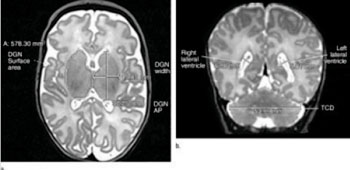
Image: (a) Axial T2-weighted image shows deep gray nuclei (DGN) width, anteroposterior (AP) distance, and surface area. (b) Coronal T2- weighted image shows lateral ventricle atrial measures and transverse cerebellar diameter (TCD) (Photo courtesy of the Radiological Society of North America).
Infants born 32 to 36 weeks into gestation may have smaller brains and other brain defects that could lead to long-term developmental difficulties, according to a new imaging study.
A lot of the existing information on preterm birth and brain development has been gathered from studies of individuals born very preterm, or less than 32 weeks into gestation at birth.
For the new study, published online June 2014 in the journal Radiology, researchers in Australia focused on moderate and late preterm (MLPT) infants—those born between 32 weeks, zero days, and 36 weeks, six days, into gestation. MLPT babies account for approximately 80% of all preterm births and are responsible for much of the rise in the rates of preterm births over the last 20 years. In spite of this, up to now there have been no large-scale studies published on brain alterations linked with MLPT births that may offer clues into brain-behavior relationships in this group of children.
“In those very preterm babies, brain injury from bleeding into the brain or a lack of blood flow, oxygen or nutrition to the brain may explain some of the abnormal brain development that occurs,” said the study’s lead author, Jennifer M. Walsh, MBBCh, BAO., MRCPI, from the Royal Women’s Hospital (Melbourne, Australia). “However, in some preterm babies, there may be no obvious explanation for why their brain development appears slow compared with babies born on time.”
The researchers performed magnetic resonance imaging (MRI) scanning on 199 MLPT and 50 full-term-born infants (greater than 37 weeks gestation) between 38 to 44 weeks of gestation. They searched for symptoms of brain injury and compared the size and maturation of multiple brain structures in the two groups. Whereas injury rates were similar between the two groups, MLPT births were tied to smaller brain size at full-term-equivalent age. Furthermore, MLPT infants had less developed myelination in one part of the brain and more immature gyral folding compared with full term-born controls.
The findings suggest that MLPT births may disrupt the predicted course of brain growth that would normally occur in the last two or so months in utero, according to Dr. Walsh. “Given that brain growth is very rapid in the last one-third of pregnancy, it is perhaps not surprising that being born during this potentially vulnerable period may disrupt brain development,” she said.
The researchers are hoping to better determine the impact that moderate to late preterm birth has on the brain, so that they can then begin to try different treatments designed to enhance brain function and long-term outcome in these infants. “Medications, along with early intervention to help parents understand their baby’s needs, have been effective in helping very preterm babies catch up to their term-born peers,” Dr. Walsh said. “However, whether any of the existing treatments will help babies born between 32 and 36 weeks is unknown, as they have not been studied very much at all.”
The researchers plan to follow the infants in the study group through childhood to learn more about the relationship between brain abnormalities and later outcomes. They also are evaluating additional MRI information about brain structure and function in these children. “Understanding what problems they have and what might be causing them is the first step in trying to improve their long-term outcome,” Dr. Walsh said.
Related Links:
Royal Women’s Hospital





A lot of the existing information on preterm birth and brain development has been gathered from studies of individuals born very preterm, or less than 32 weeks into gestation at birth.
For the new study, published online June 2014 in the journal Radiology, researchers in Australia focused on moderate and late preterm (MLPT) infants—those born between 32 weeks, zero days, and 36 weeks, six days, into gestation. MLPT babies account for approximately 80% of all preterm births and are responsible for much of the rise in the rates of preterm births over the last 20 years. In spite of this, up to now there have been no large-scale studies published on brain alterations linked with MLPT births that may offer clues into brain-behavior relationships in this group of children.
“In those very preterm babies, brain injury from bleeding into the brain or a lack of blood flow, oxygen or nutrition to the brain may explain some of the abnormal brain development that occurs,” said the study’s lead author, Jennifer M. Walsh, MBBCh, BAO., MRCPI, from the Royal Women’s Hospital (Melbourne, Australia). “However, in some preterm babies, there may be no obvious explanation for why their brain development appears slow compared with babies born on time.”
The researchers performed magnetic resonance imaging (MRI) scanning on 199 MLPT and 50 full-term-born infants (greater than 37 weeks gestation) between 38 to 44 weeks of gestation. They searched for symptoms of brain injury and compared the size and maturation of multiple brain structures in the two groups. Whereas injury rates were similar between the two groups, MLPT births were tied to smaller brain size at full-term-equivalent age. Furthermore, MLPT infants had less developed myelination in one part of the brain and more immature gyral folding compared with full term-born controls.
The findings suggest that MLPT births may disrupt the predicted course of brain growth that would normally occur in the last two or so months in utero, according to Dr. Walsh. “Given that brain growth is very rapid in the last one-third of pregnancy, it is perhaps not surprising that being born during this potentially vulnerable period may disrupt brain development,” she said.
The researchers are hoping to better determine the impact that moderate to late preterm birth has on the brain, so that they can then begin to try different treatments designed to enhance brain function and long-term outcome in these infants. “Medications, along with early intervention to help parents understand their baby’s needs, have been effective in helping very preterm babies catch up to their term-born peers,” Dr. Walsh said. “However, whether any of the existing treatments will help babies born between 32 and 36 weeks is unknown, as they have not been studied very much at all.”
The researchers plan to follow the infants in the study group through childhood to learn more about the relationship between brain abnormalities and later outcomes. They also are evaluating additional MRI information about brain structure and function in these children. “Understanding what problems they have and what might be causing them is the first step in trying to improve their long-term outcome,” Dr. Walsh said.
Related Links:
Royal Women’s Hospital

Gold Member
Blood Gas Analyzer
i-Check200
Tourniquet System
heidi– mein Tourniquet
Hypodermic Syringe
SurTract™ Safety Syringe
Latest Critical Care News
- Anatomical Guidance System Improves Trauma Ultrasound Acquisition by Novice Users
- AI-Enhanced Handheld Ultrasound Improves Carotid Plaque Detection
- Noninvasive Imaging Approach Aims to Detect Basal Cell Carcinoma Before It Becomes Visible
- New Sensor Could Bring Contactless Vital Sign Monitoring to Hospital Bedsides
- Diamond-Based Magnetometers Could Enable New Approaches to Heart Monitoring
- Emergency Department Campaign Reduces CT Use Without Identified Missed Injuries
- Portable Artificial Lung Technology Could Reshape Care for Chronic Lung Disease
- FDA Clears Multi-Device ECG Analysis Software for Extended Monitoring
- Multimodal AI Improves Treatment Response Prediction in Lung Cancer
- New Approach Helps Identify Materials for More Reliable Wearable Electronics
- Scotland Adopts Respiratory Infection Control Guidance Informed by Aerosol Research
- Hospital Safety Program Linked to 65% Reduction in MRSA Bloodstream Infections
- Wearable Microneedle Device Aims to Enable Remotely Managed Pain Relief
- Octopus-Inspired Patch Improves Adhesion for Transdermal Drug Delivery
- Multimodal AI System Tracks Early Warning Signs of Mental Health Crisis
- Self-Folding Paper Sensor Conforms to Body Without Straps or Adhesives
Channels
Artificial Intelligence
view channel
AI Improves Non-Contrast CT Interpretation for Time-Sensitive Stroke Assessment
Acute ischemic stroke occurs when a blood vessel in the brain becomes blocked, requiring rapid diagnosis to enable timely reperfusion therapy. Emergency departments often use computed tomography angiography... Read more
AI-Powered ECG Model Gains FDA Authorization for Emergency Cardiac Assessment
Powerful Medical’s PMcardio Queen of Hearts (ACS AI ECG Model) has received U.S. Food and Drug Administration (FDA) De Novo authorization, announced on September 15, 2026. The model is designed to identify... Read more
Critical Care
view channel
Anatomical Guidance System Improves Trauma Ultrasound Acquisition by Novice Users
Focused assessment with sonography for trauma (FAST) helps clinicians detect free fluid and signs of internal bleeding in injured patients, but obtaining reliable images can be difficult for inexperienced operators.... Read more
AI-Enhanced Handheld Ultrasound Improves Carotid Plaque Detection
Handheld ultrasound can miss small or faint carotid plaque, creating uncertainty in community screening. This gap makes it harder for frontline clinicians to decide who needs confirmatory imaging, closer... Read more
Noninvasive Imaging Approach Aims to Detect Basal Cell Carcinoma Before It Becomes Visible
Basal cell carcinoma is the most common form of skin cancer and can damage nearby structures such as the nose or eyes when it develops on the face. Diagnosis typically depends on visible skin changes,... Read more
Surgical Techniques
view channel
Light-Activated Nanoparticles May Offer New Approach to Retinal Prostheses
Retinitis pigmentosa is a degenerative retinal disorder in which photoreceptors progressively die, reducing visual signals to the brain while leaving surviving inner retinal circuits underused.... Read more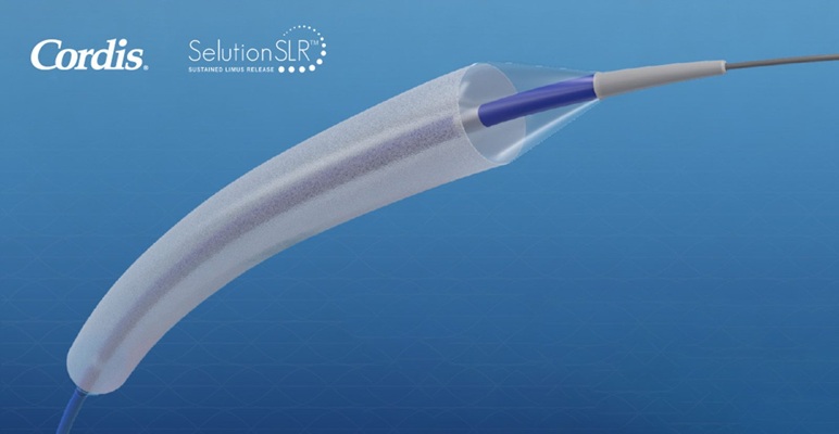
FDA Approves First Sirolimus Drug-Eluting Balloon for Coronary In-Stent Restenosis
Coronary in-stent restenosis (ISR) accounts for about 10% of the nearly one million percutaneous coronary interventions performed annually in the United States. Treating recurrent narrowing without another... Read more
Patient Care
view channel
EHR Model Flags High-Risk Periods in Patients with Metastatic Breast Cancer
Determining when patients with metastatic breast cancer are nearing the end of life remains difficult, often leading to intensive interventions with limited benefit. Prognostic uncertainty can delay goals-of-care... Read more
Virtual Reality Helps Claustrophobic Patients Complete Cardiac PET/CT Scans
Anxiety and claustrophobia—intense fear of confined spaces—often prevent patients from completing cardiac positron emission tomography/computed tomography (PET/CT). Aborted scans delay diagnosis, consume... Read more

Health IT
view channel
New Tablet Platform Extends Stroke Neurorehabilitation Beyond the Clinic
Continuity and intensity of neurorehabilitation after stroke are critical for functional recovery, but access to ongoing therapy at home often remains limited. Hospitals and outpatient programs therefore... Read more
Smartphone App Improves Blood Pressure Control in Hypertension
Hypertension, or high blood pressure, is a major modifiable driver of cardiovascular, renal, and cerebrovascular morbidity. Despite effective therapies, many adults fail to achieve control, leading to... Read more

Point of Care
view channel
Continuous Glucose Monitoring Identifies Cardiometabolic Risk in Adults Without Diabetes
Dysglycemia—abnormal blood glucose regulation—can fluctuate throughout the day and often escape conventional screening. Clinicians typically rely on fasting plasma glucose and hemoglobin A1c, which offer... Read more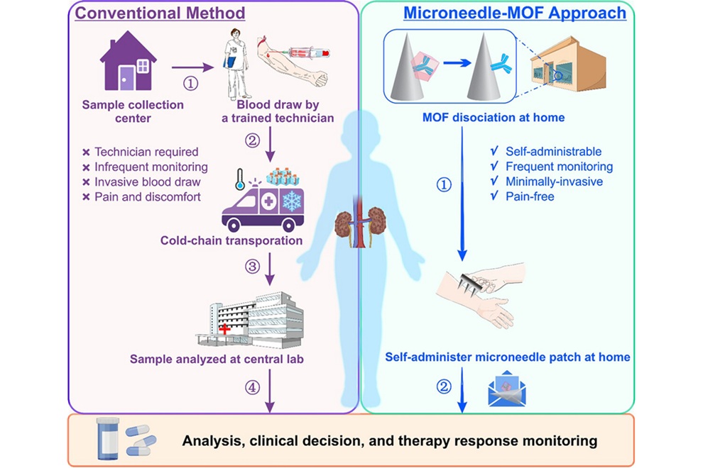
Microneedle Patch Enables At-Home Monitoring of Acute Kidney Injury
Kidney disease is often silent in its early stages, making timely detection difficult outside clinical settings. Early biomarkers such as neutrophil gelatinase–associated lipocalin can require blood draws... Read more

Business
view channel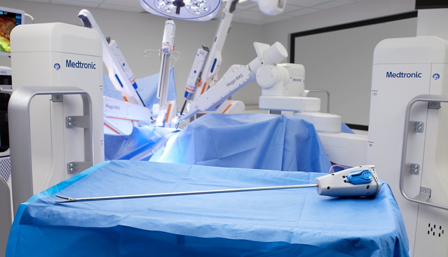
Medtronic Receives FDA Clearance for Vessel-Sealing Instrument for Robotic Surgery
As robotic-assisted surgery expands across U.S. hospitals, teams increasingly seek energy instruments with the familiarity and performance of tools used in open and laparoscopic procedures.... Read more
Robotic Single-Port System Gains CE Mark for Transvaginal Gynecologic Procedures
Intuitive (Sunnyvale, CA, USA) announced that it has received CE mark approval for use of the da Vinci SP Single Port surgical system in transvaginal gynecologic procedures, marking the first such indication... Read more