We use cookies to understand how you use our site and to improve your experience. This includes personalizing content and advertising. To learn more, click here. By continuing to use our site, you accept our use of cookies. Cookie Policy.

HospiMedica

Download Mobile App
Recent News AI Critical Care Surgical Techniques Patient Care Medical Imaging Health IT Point of Care Business Focus

Growth Guidance System Straightens Spinal Deformities

By HospiMedica International staff writers
Posted on 10 Sep 2014
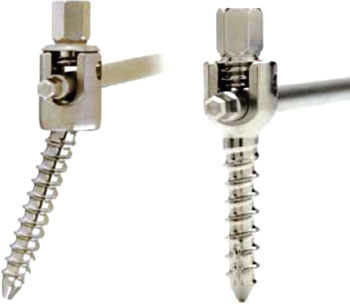
Medtronic\'s SHILLA multi-axial screw
The SHILLA multi-axial screw (left) and fixed angle screw (right) (Photo courtesy of Medtronic)
A novel growth guidance system treats skeletally immature pediatric patients diagnosed with severe early-onset spinal deformities.

The SHILLA Growth Guidance System is intended for use in pediatric patients with a severe, progressive, life-threatening, scoliosis—defined as a greater than 40° coronal plane Cobb angle or a rib-vertebral angle difference greater than 20°—associated with thoracic and spinal insufficiency that requires surgical treatment before 10 years of age. The system maintains alignment correction and stability during growth, allowing the natural increase of trunk height and unimpeded development of the thoracic cavity. The system is intended to be removed after skeletal maturity.

A unique non-locking set screw at the proximal and distal portions of the construct's rods allow the rod to slide through the screw heads as the child's spine grows, while still providing correction of the deformity. The technology allows correction while maintaining alignment over time, minimizing the need for periodic lengthening procedures and offering effective management of spinal curvature while still harnessing the child's natural growth. The SHILLA Growth Guidance System is a product of Medtronic (Minneapolis, MN, USA), and has been approved by the US Food and Drug Administration (FDA).

“Early onset scoliosis is extremely difficult to treat. The current gold standard technique to manage scoliosis long-term is to fuse the spine, but in children who are still growing this can have serious complications,” said SHILLA inventor orthopedic surgeon Richard McCarthy, MD, of Arkansas Children's Hospital (Little Rock, USA). “Until now we were only able to offer operations which use implants to stabilize the curve in the spine, but these frequently mean twice-yearly surgeries as a child grows.”

Scoliosis is a medical condition in which the spine is curved from side to side. Although a complex three-dimensional deformity, on an X-ray the spine of an individual with scoliosis may look more like an "S" or a "C" than a straight line. Scoliosis is typically classified as congenital, idiopathic, or neuromuscular, when it has developed as a secondary symptom of another condition, such as spina bifida, cerebral palsy, spinal muscular atrophy, or physical trauma.

Related Links:

Medtronic
Arkansas Children's Hospital


Gold Member
STI Test
Vivalytic Sexually Transmitted Infection (STI) Array
Radiology Monitor
MDNC-6121 Barco Nio Color 5.8MP
Patient Preoperative Skin Preparation
BD ChloraPrep
Desk Aneroid Sphyg
Diagnostix 750D+
Read the full article by registering today, it's FREE! It's Free!
Register now for FREE to HospiMedica.com and get access to news and events that shape the world of Hospital Medicine.
  • Free digital version edition of HospiMedica International sent by email on regular basis
  • Free print version of HospiMedica International magazine (available only outside USA and Canada).
  • Free and unlimited access to back issues of HospiMedica International in digital format
  • Free HospiMedica International Newsletter sent every week containing the latest news
  • Free breaking news sent via email
  • Free access to Events Calendar
  • Free access to LinkXpress new product services
  • REGISTRATION IS FREE AND EASY!
Click here to Register


Channels

Medical Imaging

view channel
Images from patient T1, who had menstrual cycle–dependent right shoulder pain. (A) Maximum-intensity-projection images show abnormal findings for right diaphragm (arrowhead), bilateral round ligaments, peritoneum around bilateral ovaries, and left fallopian tube. Combined PET/MRI show hyperintense lesion with focal uptake inferior of right diaphragm, indicative of endometriosis (arrowhead, B). Confirmatory laparoscopy demonstrated extensive pelvic disease and implants of right diaphragm (C) that stained intensely positive for FAP (D).  (Image Credit: Schindler P, Brandt J, Bobe S, et al. Initial results of FAPI PET/MRI to assess the extent of endometriosis. J Nucl Med. 2026;67(8):1232–1238. doi:10.2967/jnumed.125.271376)

Targeted PET/MRI Improves Detection and Preoperative Mapping of Endometriosis

Endometriosis is a chronic inflammatory condition in which endometrial-like tissue grows outside the uterus, causing pelvic pain, infertility, and reduced quality of life. Conventional imaging can underestimate... Read more

Business

view channel
Image: Sempresto’s Smartphone-Integrated Epinephrine Auto-Injector Wins Red Dot Design Award (Photo courtesy of Sempresto)

Smartphone-Integrated Epinephrine Auto-Injector Concept Wins Red Dot Design Award

Severe allergic reactions can escalate rapidly and require prompt epinephrine, yet many at-risk patients do not consistently carry their auto-injector. With food allergies affecting an estimated 220 million... Read more