


Imaging Shows Asymptomatic Atherosclerosis Linked to Cognitive Impairment
|
By HospiMedica International staff writers Posted on 08 Dec 2014 |


|
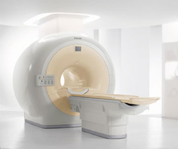
Image: The Philips Healthcare Achieva MRI system (Photo courtesy of RSNA).
In an imaging study of nearly 2,000 adults, researchers found that an accumulation of plaque in the body’s major arteries was linked with mild cognitive impairment.
The study’s findings were conducted at the University of Texas (UT) Southwestern Medical Center (Dallas, USA) and presented December 2014 at the annual meeting of the Radiological Society of North America (RSNA), held in Chicago (IL, USA). “It is well established that plaque buildup in the arteries is a predictor of heart disease, but the relationship between atherosclerosis and brain health is less clear,” said Christopher D. Maroules, MD, a radiology resident at UT Southwestern Medical Center in Dallas. “Our findings suggest that atherosclerosis not only affects the heart but also brain health.”
Atherosclerosis is a disorder in which cholesterol, fat, and other substances gather in the arteries, forming a substance called plaque that can build up, restricting blood flow. It can occur in any artery of the body, including the carotid, which supplies blood to the brain, coronary arteries and the aorta, which carries oxygenated blood from the heart through the abdomen to the rest of body.
In the study, researchers analyzed the test results of 1,903 participants (mean age, 44 years) in the Dallas Heart Study, a multiethnic population-based study of adults from Dallas County, Texas. The participants included both men and women who had no symptoms of cardiovascular disease. Study participants completed the Montreal Cognitive Assessment (MoCA), a 30-point standardized test for detecting mild cognitive impairment, and underwent magnetic resonance imaging (MRI) of the brain to identify white matter hyperintensity (WMH) volume. Bright white spots known as high signal intensity areas on a brain MR images indicate abnormal changes within the white matter. “Increased white matter hyperintensity volume is part of the normal aging process,” Dr. Maroules explained. “But excessive WMH volume is a marker for cognitive impairment.”
Study participants also underwent imaging exams to measure the buildup of plaque in the arteries in three distinct vascular areas of the body: MRI to measure wall thickness in the carotid arteries and the abdominal aorta, and computed tomography (CT) to measure coronary artery calcium, or the amount of calcified plaque in the arteries of the heart.
Using the findings, researchers performed a statistical regression to correlate the incidence of atherosclerosis and mild cognitive impairment. After adjusting for traditional risk factors for atherosclerosis, including age, ethnicity, male sex, diabetes, hypertension, smoking, and body mass index, they found independent relationships between atherosclerosis in all three vascular areas of the body and cognitive health, as measured by MoCA scores and white matter hyperintensity volume on MR images.
Individuals in the highest quartile of internal carotid wall thickness were 21% more likely to have cognitive impairment as measured by a low MoCA score. An increasing coronary artery calcium score was predictive of large white matter intensity volume on MRI. “These results underscore the importance of identifying atherosclerosis in its early stages, not just to help preserve heart function, but also to preserve cognition and brain health,” Dr. Maroules said.
Dr. Maroules noted that the MRI and CT imaging techniques provide valuable prognostic information about an individual’s downstream health risks. “Plaque buildup in blood vessels throughout the body offers us a window into brain health,” he said. “Imaging with CT and MRI has an important role in identifying patients who are at a higher risk for cognitive impairment.”
RSNA (Oak Brook, IL, USA) is an association of more than 54,000 radiologists, radiation oncologists, medical physicists and related scientists, encouraging excellence in patient care and health care delivery through education, research and technologic advances.
Related Links:
University of Texas Southwestern Medical Center





The study’s findings were conducted at the University of Texas (UT) Southwestern Medical Center (Dallas, USA) and presented December 2014 at the annual meeting of the Radiological Society of North America (RSNA), held in Chicago (IL, USA). “It is well established that plaque buildup in the arteries is a predictor of heart disease, but the relationship between atherosclerosis and brain health is less clear,” said Christopher D. Maroules, MD, a radiology resident at UT Southwestern Medical Center in Dallas. “Our findings suggest that atherosclerosis not only affects the heart but also brain health.”
Atherosclerosis is a disorder in which cholesterol, fat, and other substances gather in the arteries, forming a substance called plaque that can build up, restricting blood flow. It can occur in any artery of the body, including the carotid, which supplies blood to the brain, coronary arteries and the aorta, which carries oxygenated blood from the heart through the abdomen to the rest of body.
In the study, researchers analyzed the test results of 1,903 participants (mean age, 44 years) in the Dallas Heart Study, a multiethnic population-based study of adults from Dallas County, Texas. The participants included both men and women who had no symptoms of cardiovascular disease. Study participants completed the Montreal Cognitive Assessment (MoCA), a 30-point standardized test for detecting mild cognitive impairment, and underwent magnetic resonance imaging (MRI) of the brain to identify white matter hyperintensity (WMH) volume. Bright white spots known as high signal intensity areas on a brain MR images indicate abnormal changes within the white matter. “Increased white matter hyperintensity volume is part of the normal aging process,” Dr. Maroules explained. “But excessive WMH volume is a marker for cognitive impairment.”
Study participants also underwent imaging exams to measure the buildup of plaque in the arteries in three distinct vascular areas of the body: MRI to measure wall thickness in the carotid arteries and the abdominal aorta, and computed tomography (CT) to measure coronary artery calcium, or the amount of calcified plaque in the arteries of the heart.
Using the findings, researchers performed a statistical regression to correlate the incidence of atherosclerosis and mild cognitive impairment. After adjusting for traditional risk factors for atherosclerosis, including age, ethnicity, male sex, diabetes, hypertension, smoking, and body mass index, they found independent relationships between atherosclerosis in all three vascular areas of the body and cognitive health, as measured by MoCA scores and white matter hyperintensity volume on MR images.
Individuals in the highest quartile of internal carotid wall thickness were 21% more likely to have cognitive impairment as measured by a low MoCA score. An increasing coronary artery calcium score was predictive of large white matter intensity volume on MRI. “These results underscore the importance of identifying atherosclerosis in its early stages, not just to help preserve heart function, but also to preserve cognition and brain health,” Dr. Maroules said.
Dr. Maroules noted that the MRI and CT imaging techniques provide valuable prognostic information about an individual’s downstream health risks. “Plaque buildup in blood vessels throughout the body offers us a window into brain health,” he said. “Imaging with CT and MRI has an important role in identifying patients who are at a higher risk for cognitive impairment.”
RSNA (Oak Brook, IL, USA) is an association of more than 54,000 radiologists, radiation oncologists, medical physicists and related scientists, encouraging excellence in patient care and health care delivery through education, research and technologic advances.
Related Links:
University of Texas Southwestern Medical Center

Gold Member
Neonatal Heel Incision Device
Tenderfoot
Patient Preoperative Skin Preparation
BD ChloraPrep
Hypodermic Syringe
SurTract™ Safety Syringe
Latest Critical Care News
- Scotland Adopts Respiratory Infection Control Guidance Informed by Aerosol Research
- Hospital Safety Program Linked to 65% Reduction in MRSA Bloodstream Infections
- Wearable Microneedle Device Aims to Enable Remotely Managed Pain Relief
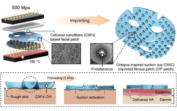
- Octopus-Inspired Patch Improves Adhesion for Transdermal Drug Delivery
- Multimodal AI System Tracks Early Warning Signs of Mental Health Crisis
- Self-Folding Paper Sensor Conforms to Body Without Straps or Adhesives
- Patch-Based Therapy Promotes Myelin Repair in Multiple Sclerosis
- Rapid EHR Surveillance Helps Protect High-Risk Patients During Heat Waves
- Remote Cardiac Device Monitoring System Shows Safety and Cost Benefits
- Ambulatory ECG Monitor Improves Arrhythmia Detection in Syncope
- New Prediction Tool Supports Confident Prescribing of Heart Failure Medicines
- Machine Learning Models Identify Higher-Risk Pregnancies Earlier in Care
- Wearable Hormone Monitor Enables At-Home Detection of Primary Aldosteronism
- Contactless AI Tool Detects Hypertension and Diabetes from Facial Video
- AI Tool Predicts Bladder Cancer from Electronic Health Records
- Personalized Neuromodulation System Receives FDA Clearance for PTSD Treatment
Channels
Artificial Intelligence
view channel
Explainable AI Tool Identifies High-Risk Patients Before Heart Attack Reperfusion
Intramyocardial hemorrhage is bleeding within the heart muscle that can occur after blood flow is restored during heart attack treatment. It affects about 40% of patients treated for ST-segment elevation... Read more
AI Tools Show Potential to Detect Subtle Signs of Interval Breast Cancer
Interval breast cancers, diagnosed after a negative screening mammogram and before the next scheduled exam, remain an important measure of screening performance and are often more aggressive.... Read more

Critical Care
view channel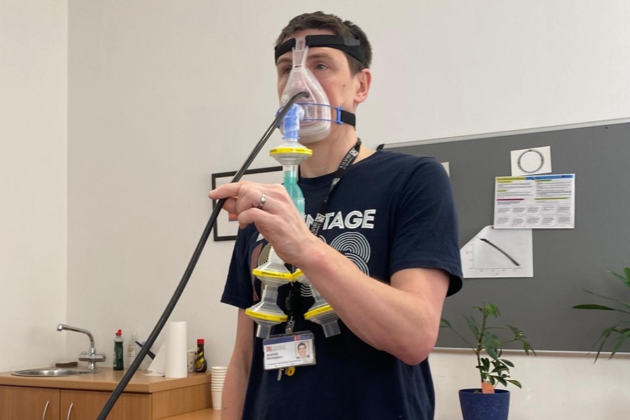
Scotland Adopts Respiratory Infection Control Guidance Informed by Aerosol Research
Preventing respiratory infection transmission in hospitals remains a core patient and staff safety priority. Many precautions have hinged on the assumption that certain clinical interventions create high-risk... Read more
Hospital Safety Program Linked to 65% Reduction in MRSA Bloodstream Infections
Hospital-onset methicillin-resistant Staphylococcus aureus (MRSA) bloodstream infections are difficult to treat and increase morbidity, mortality, and costs for acutely ill patients. Vulnerability rises... Read more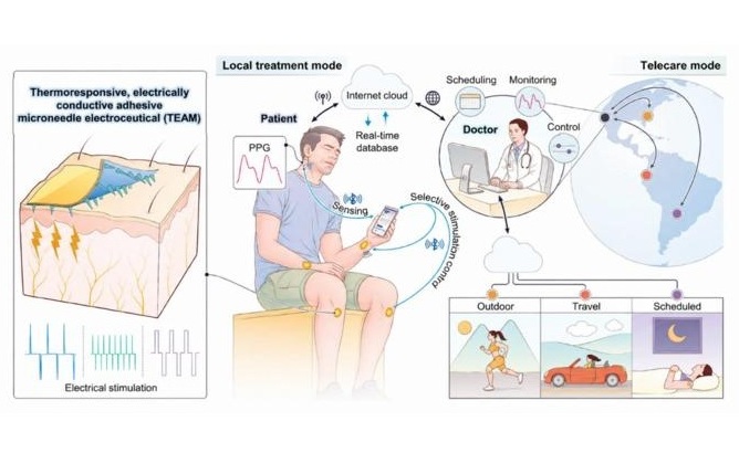
Surgical Techniques
view channel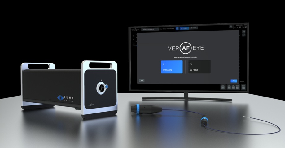
FDA Clears Integrated Platform for Real-Time Cardiac Imaging and Catheter Guidance
Catheter-based treatment of cardiac arrhythmias relies on precise, real-time visualization and navigation for safe, accurate therapy delivery. Yet electrophysiology teams often use separate imaging, mapping,... Read more
New Microcatheter Enhances Lesion Crossing in Complex Coronary Interventions
Crossing resistant coronary lesions during percutaneous coronary intervention can be hindered by tortuous or calcified anatomy, delaying therapy and complicating device delivery. Many microcatheters rely... Read more
Integrated Perioperative Care Model Improves Recovery in Older Surgical Patients
Older adults undergoing major surgery often experience anxiety and depression that can slow rehabilitation and worsen functional recovery. Many also take central nervous system–active medications that... Read more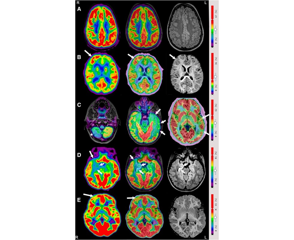
Integrated PET/MRI and EEG Improves Presurgical Lesion Localization in Pediatric Focal Epilepsy
Focal epilepsy, in which seizures arise from a discrete brain region, requires precise lesion localization before surgery. Many children have negative or inconclusive magnetic resonance imaging (MRI),... Read morePatient Care
view channel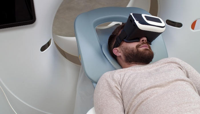
Virtual Reality Helps Claustrophobic Patients Complete Cardiac PET/CT Scans
Anxiety and claustrophobia—intense fear of confined spaces—often prevent patients from completing cardiac positron emission tomography/computed tomography (PET/CT). Aborted scans delay diagnosis, consume... Read more
Weighted Blankets Reduce Anxiety in Hospitalized Adults
Anxiety is common among hospitalized adults and can impair recovery, disrupt sleep, and worsen mental health. Although clinicians often use nonpharmacologic strategies, bedside options that act quickly... Read more
Health IT
view channel
Registry-Driven Digital Outreach Boosts Use of Heart Failure Medicines
Heart failure affects more than 64 million people worldwide and is linked to high mortality, reduced quality of life, and heavy healthcare use. Although sodium-glucose cotransporter 2 inhibitors are proven... Read more
Nurse-Coordinated Care Model Improves Heart Failure Outcomes
Heart failure imposes heavy mortality, morbidity, and cost burdens worldwide, yet proven therapies remain underused in routine practice. This inadequate uptake contributes to preventable hospitalizations... Read more

Point of Care
view channel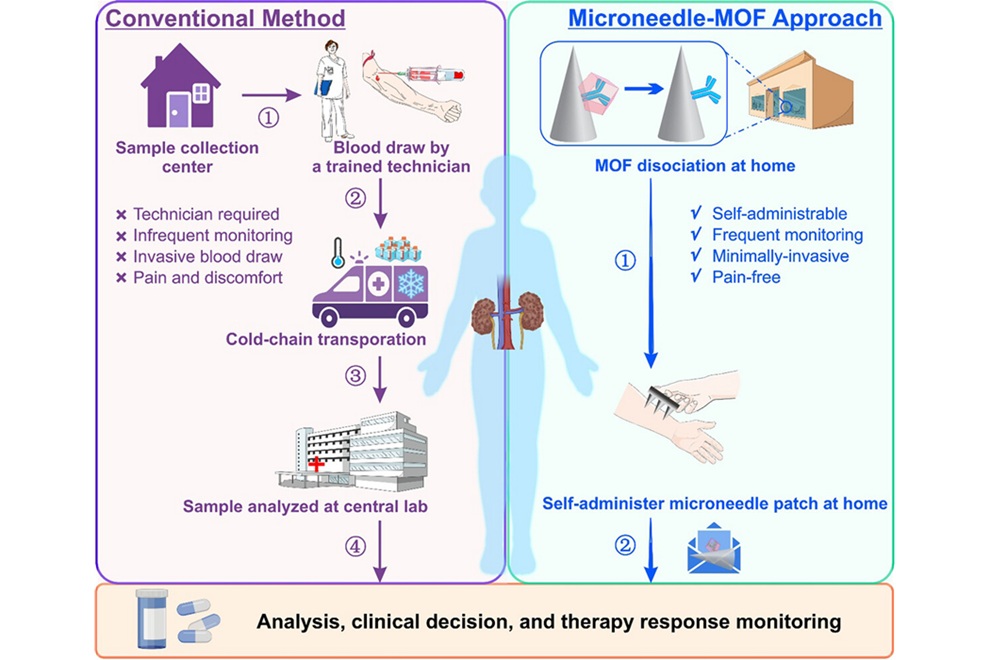
Microneedle Patch Enables At-Home Monitoring of Acute Kidney Injury
Kidney disease is often silent in its early stages, making timely detection difficult outside clinical settings. Early biomarkers such as neutrophil gelatinase–associated lipocalin can require blood draws... Read more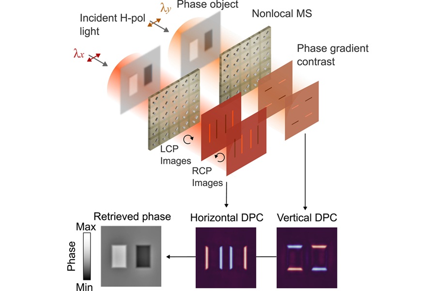
Ultrathin Metasurface Could Bring Quantitative Phase Imaging to Portable Devices
Point-of-care imaging often depends on bulky optical systems that have difficulty visualizing nearly transparent cells and tissues without staining. These limitations can slow diagnosis and restrict use... Read more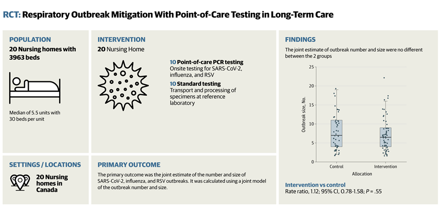
Business
view channel
Robotic Single-Port System Gains CE Mark for Transvaginal Gynecologic Procedures
Intuitive (Sunnyvale, CA, USA) announced that it has received CE mark approval for use of the da Vinci SP Single Port surgical system in transvaginal gynecologic procedures, marking the first such indication... Read more