Dissolvable Surgical Clip Improves Diagnostic Imaging
|
By HospiMedica International staff writers Posted on 13 May 2015 |
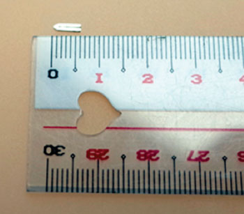
Image: The Kobe dissolvable surgical clip (Photo courtesy of Kobe University).
A safe surgical clip could reduce postoperative complication rates and minimize problems associated with diagnostic imaging.
Developed by researchers at Kobe University (Japan), the 5-mm dissolvable surgical clip is made of a magnesium alloy that dissolves and is absorbed by the body after a certain period of time. The alloy also contains calcium and zinc to improve its microstructure, ensuring fastening ability and formability. To evaluate the safety of the clip, an implantation study was conducted in a subcutaneous mouse model. The results showed that very little gas was produced as the clip dissolved, and there was no inflammation of the surrounding tissues after 12 weeks.
As the volume of the implanted clip was reduced by almost half during that time, the researchers concluded it would likely dissolve completely within one year. Blood tests revealed that levels of magnesium and other substances in the blood were in the normal range after 12 weeks. To evaluate the functionality of the clip, it was tested in a rat model in which the biliary duct, portal vein, hepatic artery, and hepatic vein were occluded with the clip and a partial liver was removed. During a monitoring period of eight weeks, the clip functioned properly, and micro CT scanning revealed that the quality of images was not degraded.
“We will conduct further in vivo studies and a clinical study within two to three years,” said metallurgical engineer, Prof. Toshiji Mukai, PhD, of the Kobe University Graduate School of Engineering, who was involved in developing the clip. “Kobe University works toward the development of new medical devices. We will continue to promote collaboration between the graduate schools of medicine and engineering.”
Most surgical clips are made of titanium, and as many as 30 to 40 clips may be used during a single surgical procedure. The retained clips lead to diminished quality of computed tomography (CT) and magnetic resonance imaging (MRI) images around the wound area, and can potentially cause complications.
Related Links:
Kobe University
Latest Critical Care News
- Injectable Disease-Fighting Nanorobots to Improve Precision Cancer Therapy
- Web-Based Tool Enables Early Detection and Prevention of Chronic Kidney Disease
- Tiny Sensor to Transform Head Injury Detection
- Bacterial Behavior Breakthrough to Improve Infection Prevention in Biomedical Devices
- Implanted 'Living Skin' Indicates Internal Inflammation Without Blood Samples
- AI Tool Improves Speed and Accuracy of Cervical Cancer Treatment Planning
- Ultrasonic Sensor Enables Cuffless and Non-Invasive Blood Pressure Measurement
- Simple Change in Sepsis Treatment Could Save Thousands of Lives
- AI-Powered ECG Analysis Enables Early COPD Detection
- Soft Wireless Implant Treats Inflammatory Bowel Disease
- Pill Reports from Stomach When It Has Been Swallowed
- Wireless Sensing Technology Enables Touch-Free Diagnostics of Common Lung Diseases
- Early Detection and Targeted Blood Purification Could Prevent Kidney Failure in ICU Patients
- New Cancer Treatment Uses Sound-Responsive Particles to Soften Tumors
- Sprayable Powder-Type Hemostatic Agent Stops Bleeding in One Second
- Ultra-Stable Mucus-Inspired Hydrogel Boosts Gastrointestinal Wound Healing
Channels
Artificial Intelligence
view channelSurgical Techniques
view channel
Ultrasound Device Offers Non-Invasive Treatment for Kidney Stones
The U.S. Food and Drug Administration has granted 510(k) clearance to SonoMotion’s Break Wave lithotripsy device, which fragments stones non-invasively with focused ultrasound and requires no anesthesia.... Read more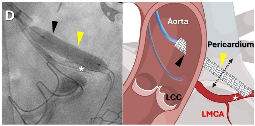
Minimally Invasive Coronary Artery Bypass Method Offers Safer Alternative to Open-Heart Surgery
Coronary artery obstruction is a rare but often fatal complication of heart-valve replacement, particularly in patients with complex anatomy or prior cardiac interventions. In such cases, traditional open-heart... Read morePatient Care
view channel
Revolutionary Automatic IV-Line Flushing Device to Enhance Infusion Care
More than 80% of in-hospital patients receive intravenous (IV) therapy. Every dose of IV medicine delivered in a small volume (<250 mL) infusion bag should be followed by subsequent flushing to ensure... Read more
VR Training Tool Combats Contamination of Portable Medical Equipment
Healthcare-associated infections (HAIs) impact one in every 31 patients, cause nearly 100,000 deaths each year, and cost USD 28.4 billion in direct medical expenses. Notably, up to 75% of these infections... Read more
Portable Biosensor Platform to Reduce Hospital-Acquired Infections
Approximately 4 million patients in the European Union acquire healthcare-associated infections (HAIs) or nosocomial infections each year, with around 37,000 deaths directly resulting from these infections,... Read moreFirst-Of-Its-Kind Portable Germicidal Light Technology Disinfects High-Touch Clinical Surfaces in Seconds
Reducing healthcare-acquired infections (HAIs) remains a pressing issue within global healthcare systems. In the United States alone, 1.7 million patients contract HAIs annually, leading to approximately... Read moreHealth IT
view channel
EMR-Based Tool Predicts Graft Failure After Kidney Transplant
Kidney transplantation offers patients with end-stage kidney disease longer survival and better quality of life than dialysis, yet graft failure remains a major challenge. Although a successful transplant... Read more
Printable Molecule-Selective Nanoparticles Enable Mass Production of Wearable Biosensors
The future of medicine is likely to focus on the personalization of healthcare—understanding exactly what an individual requires and delivering the appropriate combination of nutrients, metabolites, and... Read moreBusiness
view channel
WHX in Dubai (formerly Arab Health) to bring together key UAE government entities during the groundbreaking 2026 edition
World Health Expo (WHX), formerly Arab Health, will bring together the UAE’s health authorities and leading healthcare sector bodies when the exhibition debuts at the Dubai Exhibition Centre (DEC) from... Read more
Interoperability Push Fuels Surge in Healthcare IT Market
Hospitals still struggle to reconcile data scattered across electronic health records, laboratory systems, and billing platforms, undermining care coordination and operational efficiency.... Read more