



Radiation-Free System Helps Diagnose Scoliosis 
|
By HospiMedica International staff writers Posted on 09 Jun 2015 |


|
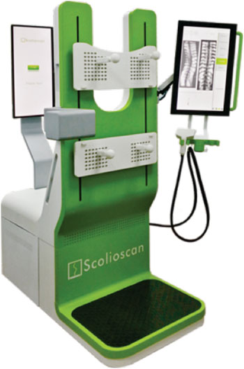
Image: The Scolioscan ultrasound scoliosis assessment system (Photo courtesy Hong Kong Polytechnic University).
Novel ultrasound technology captures the spine's three-dimensional (3D) profile using bony landmarks, enabling safer and more frequent screenings for scoliosis.
Developed by researchers at Hong Kong Polytechnic University University (PolyU; Hong Kong), the Scolioscan device uses ultrasound to assess a patient's spine in the standing posture and generate coronal images of spinal curvature, which can then be measured to determine the severity of the scoliosis (as determined by spinal rotation and deformity along different planes). The device is radiation-free and enables safer screening whenever needed, thus avoiding the health risks posed by radiation exposure, particularly in young patients.
The examination process takes about one minute to generate an ultrasound B-scan 3D image. This allows healthcare workers to detect adolescent idiopathic scoliosis (AIS) at an early stage, or avoid unnecessary treatments for patients with stable spinal angles. It also allows close follow-up and monitoring on the progress of spinal bracing or other treatments for AIS. The product has been commercialized by Telefield Medical Imaging (Hong Kong).
“The current verification standard is based on the Cobb's angle method, used to measure spinal deformity, but it does not always represent the true value of spinal curvature,” said Prof. Yongping Zheng, PhD, MEng. “The values of spinal curvature produced by Cobb's angle can vary by up to seven degrees. Therefore, further research is needed to find a better way to verify the angles measured by the Scolioscan or ultimately establish its own standard.”
Scoliosis is a medical condition in which a person's spine is curved from side to side. Although it is a complex 3D deformity, on an X-ray, viewed from the rear, the spine of an individual with scoliosis may look more like an "S" or a "C" than a straight line. Scoliosis is typically classified as congenital, idiopathic, or neuromuscular, when it has developed as a secondary symptom of another condition, such as spina bifida, cerebral palsy, spinal muscular atrophy, or physical trauma.
Related Links:
Hong Kong Polytechnic University University
Telefield Medical Imaging






Developed by researchers at Hong Kong Polytechnic University University (PolyU; Hong Kong), the Scolioscan device uses ultrasound to assess a patient's spine in the standing posture and generate coronal images of spinal curvature, which can then be measured to determine the severity of the scoliosis (as determined by spinal rotation and deformity along different planes). The device is radiation-free and enables safer screening whenever needed, thus avoiding the health risks posed by radiation exposure, particularly in young patients.
The examination process takes about one minute to generate an ultrasound B-scan 3D image. This allows healthcare workers to detect adolescent idiopathic scoliosis (AIS) at an early stage, or avoid unnecessary treatments for patients with stable spinal angles. It also allows close follow-up and monitoring on the progress of spinal bracing or other treatments for AIS. The product has been commercialized by Telefield Medical Imaging (Hong Kong).
“The current verification standard is based on the Cobb's angle method, used to measure spinal deformity, but it does not always represent the true value of spinal curvature,” said Prof. Yongping Zheng, PhD, MEng. “The values of spinal curvature produced by Cobb's angle can vary by up to seven degrees. Therefore, further research is needed to find a better way to verify the angles measured by the Scolioscan or ultimately establish its own standard.”
Scoliosis is a medical condition in which a person's spine is curved from side to side. Although it is a complex 3D deformity, on an X-ray, viewed from the rear, the spine of an individual with scoliosis may look more like an "S" or a "C" than a straight line. Scoliosis is typically classified as congenital, idiopathic, or neuromuscular, when it has developed as a secondary symptom of another condition, such as spina bifida, cerebral palsy, spinal muscular atrophy, or physical trauma.
Related Links:
Hong Kong Polytechnic University University
Telefield Medical Imaging

Gold Member
SARS‑CoV‑2/Flu A/Flu B/RSV Sample-To-Answer Test
SARS‑CoV‑2/Flu A/Flu B/RSV Cartridge (CE-IVD)
Medical Examination & Procedure Light
Vega 80
Immobilization System
Cranial 4Pi Immobilization
Latest Critical Care News
- Smart Medical Adhesive Patch Uses Heat to Enable Gentle Removal
- Smart Pulse Oximetry Supports Earlier Recognition of Arrhythmias and Respiratory Risk
- Wearable Robotic Gait Training Linked to Improved Walking Distance in ALS
- Consumer Smartwatch May Expand Access to Obstructive Sleep Apnea Screening
- Proposed Risk Score Prioritizes Sickest Patients for Heart Transplant
- Wristwatch Sensor Tracks Sodium and Potassium in Sweat in Real Time
- Smart Tear Sensors May Help Detect Shifts in Eye Health Over Time
- Wearable Smart Ring Receives FDA Clearance for Noninvasive Blood Oxygen Monitoring
- AI Tool Combining ECG and Blood Tests Reduces Unnecessary Heart Transplant Biopsies
- Deep Brain Stimulation Window May Matter for Parkinson’s Patients With GBA1 Variants
- Minimally Invasive Ultrasound-Guided Procedure Eases Carpal Tunnel Symptoms
- FDA-Approved App Enables Pulmonary Pressure Monitoring and Medication Guidance in Heart Failure
- Wearable Patch Prototype Advances Toward Integrated Automated Insulin Delivery
- Standardized Scoring Tool Supports ICU Discharge Decisions
- Battery-Free Wearable Sensor Enables Activity Recognition for Health Monitoring
- New ECG Foundation Model Enables Broad Cardiac Diagnosis and Risk Prediction
Channels
Artificial Intelligence
view channel
AI Tool Predicts Cancer Survival and Biological Age from Single Facial Photo
Assessing frailty and physiologic reserve remains challenging when clinicians consider intensive cancer treatment. Chronological age alone may not reflect how well a patient can tolerate radiation, surgery,... Read more
AI Tool Improves Smartphone Detection of Abnormal Pediatric Lung Sounds
Respiratory disease in children remains a major clinical concern worldwide. Abnormal lung sounds can indicate pediatric respiratory illness, but standard auscultation usually requires an in-person examination... Read more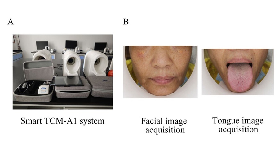

Surgical Techniques
view channel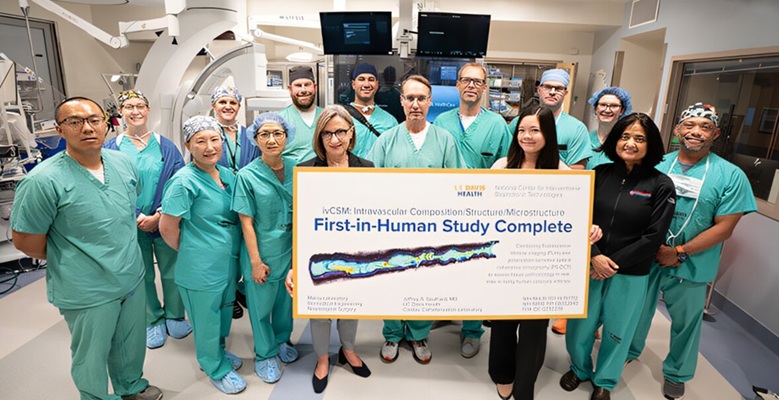
Dual-Imaging Catheter Completes Initial Human Evaluation of Coronary Plaque
Atherosclerosis involves plaque buildup in arterial walls that can narrow blood vessels and increase the risk of heart attack, stroke, and other serious cardiovascular conditions. In coronary arteries,... Read more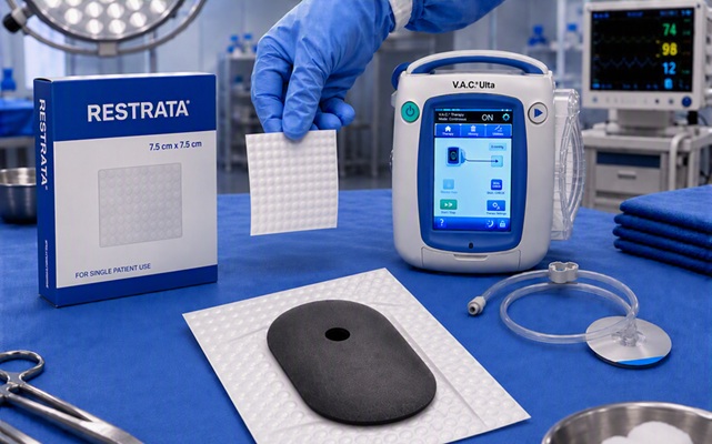
Combined Wound Healing Approach Pairs Synthetic Scaffold With Negative Pressure Therapy
Managing complex wounds requires both structural support for tissue repair and a controlled environment that protects the healing process. Frequent dressing changes can disrupt the wound bed and add complexity... Read more
Patient Care
view channel
Total-Body PET/CT Scanner Enables One-Minute Diagnostic-Quality Cancer Imaging
Positron emission tomography/computed tomography (PET/CT) combines radiotracer-based functional imaging with anatomical detail to diagnose, stage, and monitor cancer. Conventional scanners image the body... Read more
AI Platform Personalizes Support Between Visits for Cancer Survivors
Cancer survivors often manage persistent side effects after treatment, including fatigue, anxiety, uncertainty, symptoms, stress, and questions between appointments. Clinical teams need ways to maintain... Read more
At-Home Neuromodulation Shows Promise in Selected Patients With Hypertension
High blood pressure is a major risk factor for cardiovascular disease, including heart attack and stroke. Many patients receiving treatment for hypertension still remain above recommended blood pressure... Read more
EHR Model Flags High-Risk Periods in Patients with Metastatic Breast Cancer
Determining when patients with metastatic breast cancer are nearing the end of life remains difficult, often leading to intensive interventions with limited benefit. Prognostic uncertainty can delay goals-of-care... Read moreMedical Imaging
view channel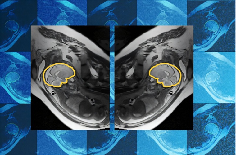
AI Tool Improves Fetal MRI for Brain Abnormality Diagnosis
Fetal brain abnormalities can be difficult to diagnose before birth when fetal motion reduces imaging quality. Routine ultrasound may flag a possible abnormality, prompting follow-up fetal MRI, but head... Read more
AI Model Detects Hidden Disease Signals in Routine CT Scans
Computed tomography (CT) scans are often performed to answer a specific diagnostic question, which can leave other potentially important findings outside the immediate clinical focus. As a result, opportunities... Read more
Routine CT Body Composition Tool Helps Identify Early Pancreatic Cancer Risk
Pancreatic ductal adenocarcinoma (PDAC), the most common type of pancreatic cancer, often goes undetected until it has spread. Surveillance programs primarily target people with inherited genetic risk... Read more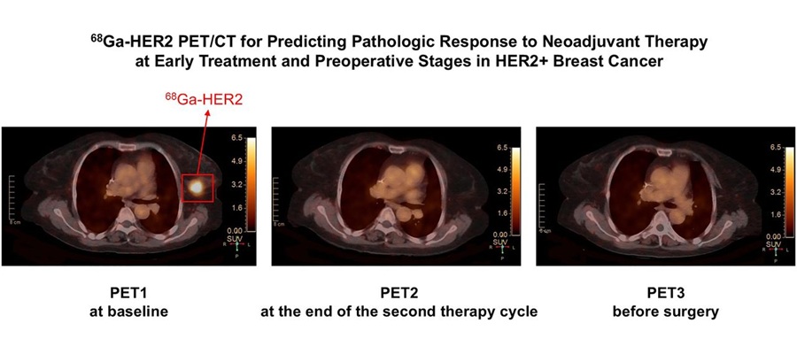
Targeted PET/CT Predicts Early Treatment Response in HER2-Positive Breast Cancer
HER2-positive breast cancer is an aggressive subtype that accounts for a substantial proportion of breast cancer cases. Patients often receive neoadjuvant therapy before surgery to shrink tumors, but assessing... Read moreHealth IT
view channel
EHR Alerts Support Dosing, Sedation, and Shock Recognition in Cardiac Care
Electronic health record alerts can add to clinician workload when they are poorly targeted. In critical cardiac care, however, delayed decisions can affect treatment timing, sedation exposure, and escalation... Read more
Adaptive Radiation QA Software Gains Validation for NeoArc Treatments
Adaptive radiation therapy allows clinicians to adjust treatment plans to daily anatomical changes such as tumor shrinkage, weight loss, or organ movement, improving precision while limiting exposure to... Read more

Point of Care
view channel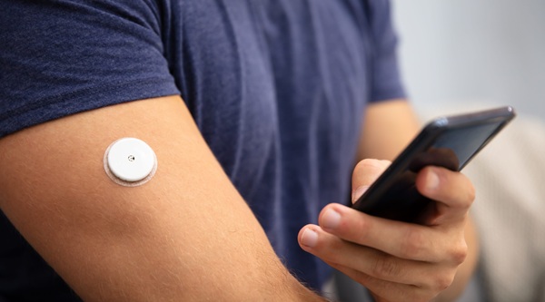
Continuous Glucose Monitoring Identifies Cardiometabolic Risk in Adults Without Diabetes
Dysglycemia—abnormal blood glucose regulation—can fluctuate throughout the day and often escape conventional screening. Clinicians typically rely on fasting plasma glucose and hemoglobin A1c, which offer... Read more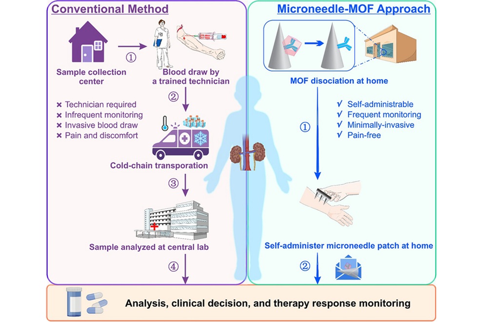
Microneedle Patch Enables At-Home Monitoring of Acute Kidney Injury
Kidney disease is often silent in its early stages, making timely detection difficult outside clinical settings. Early biomarkers such as neutrophil gelatinase–associated lipocalin can require blood draws... Read more
Business
view channel
Steris to Expand Surgical Safety Portfolio With $2.1 Billion Xodus Medical Deal
Steris has signed a definitive agreement to acquire Xodus Medical, a specialty surgical products business based in Pennsylvania, for $2.1 billion at closing. The acquisition will expand Steris’s healthcare... Read more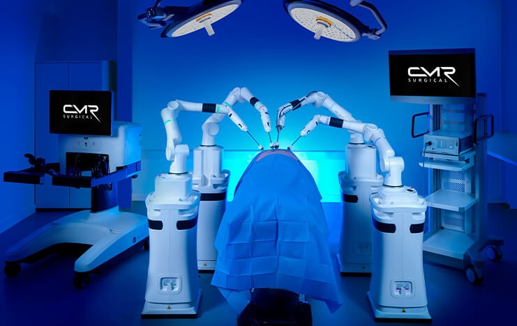
Stryker Enters Soft-Tissue Robotics Through Strategic Alliance With CMR Surgical
Stryker (Portage, MI, USA) and CMR Surgical (Cambridge, UK) have announced a strategic alliance centered on the Versius Surgical Robotic portfolio. The agreement includes an approximately USD 400 million... Read more
Terumo Neuro Acquisition Advances Liquid Embolic Technology for Chronic Subdural Hematoma
Terumo Neuro (Aliso Viejo, CA, USA) signed an agreement to acquire Arsenal Medical Inc. (Waltham, MA, USA), a privately held, clinical-stage biomaterials company. The proposed acquisition would add NeoCast,... Read more