Rapid and Automated System Analyzes Kidney Stones
|
By HospiMedica International staff writers Posted on 12 Oct 2015 |
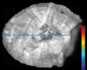
Image: A kidney stone being analyzed automatically using Raman spectrosopy (Photo courtesy of Fraunhofer IPM).
A new imaging system for rapid analysis of urinary stones immediately after the surgical procedure can help determine appropriate postoperative care.
Researchers at the Fraunhofer Institute for Physical Measurement Techniques (IPM; Freiburg, Germany) in collaboration with an industrial partner and University Medical Center Freiburg (Germany) are developing a novel Raman spectroscopy diagnostic system for rapid and automated analysis of kidney stones. The system identifies the light spectrum of the examined sample by illuminating it with a laser, identifying the singular characteristic wave spectrum via the 1% of photons reflected back.
The researchers then use computer software to filter out the fluorescent background occurring during Raman spectroscopy. The results are then compared to a spectral database that contains data on the nine pure substances that make up 99% of urinary stones, as determined by examining nearly 160 samples; the results were confirmed by conventional infrared (IR) based analysis in a reference laboratory. Since the device employs relatively inexpensive optical components, and it can work on wet, unprepared samples, the time taken to prepare specimens is substantially reduced.
“The stones previously had to be dried and pulverized prior to analysis. Our system makes this unnecessary. Stone fragments collected during the surgical procedure do not need to be further processed. They can in principle be put directly into the Raman spectrometer for analysis,” explained IPM physician and researcher Arkadiusz Miernik, MD. “Currently there are a few specialized laboratories that can carry out this procedure using large-scale analytical equipment. A compact device suitable for use in a clinical setting and allowing immediate, post interventional automated analysis is not yet available.”
“We advise stone patients to drink plenty of fluids, increase physical activities and lose weight if necessary. Unfortunately this is only a general recommendation,” added Dr. Miernik. “Once the complete system is ready for clinical use, the physician will be able to examine stone samples directly after surgical intervention on his own, thus increasing the quality of patients’ care substantially.”
Kidney stones are often no larger than a grain of rice, yet some can grow to a diameter of several centimeters, causing blockage of the ureters. If it cannot be dissolved chemically, the kidney stone is treated using extracorporeal shock-wave therapy or minimally invasive endoscopic modalities. Many of these patients suffer from disease recurrence and need retreatment, but new stone formation might be reduced by 50% if individualized follow-up care and proper measures are offered to the patient regarding dietary habits or the use of particular medication strategies, based on stone composition.
Related Links:
Fraunhofer Institute for Physical Measurement Techniques
University Medical Center Freiburg
Latest Surgical Techniques News
- Novel Endoscopy Technique Provides Access to Deep Lung Tumors
- New Study Findings Could Halve Number of Stent Procedures
- Breakthrough Surgical Device Redefines Hip Arthroscopy
- Automated System Enables Real-Time "Molecular Pathology" During Cancer Surgery
- Groundbreaking Procedure Combines New Treatments for Liver Tumors
- Ablation Reduces Stroke Risk Associated with Atrial Fibrillation
- Optical Tracking Method Identifies Target Areas in Robot-Assisted Neurosurgery
- General Anesthesia Improves Post-Surgery Outcomes for Acute Stroke Patients
- Drug-Coated Balloons Can Replace Stents Even in Larger Coronary Arteries
- Magnetic Kidney Stone Retrieval Device Outperforms Ureteroscopic Laser Lithotripsy
- Absorbable Skull Device Could Replace Traditional Metal Implants Used After Brain Surgery
- Magic Silicone Liquid Powered Robots Perform MIS in Narrow Cavities
- 'Lab-on-a-Scalpel' Provides Real-Time Surgical Insights for POC Diagnostics in OR
- Biodegradable Brain Implant Prevents Glioblastoma Recurrence
- Tiny 3D Printer Reconstructs Tissues During Vocal Cord Surgery
- Minimally Invasive Procedure for Aortic Valve Disease Has Similar Outcomes as Surgery
Channels
Critical Care
view channel
Nasal Drops Fight Brain Tumors Noninvasively
Glioblastoma is one of the most aggressive and fatal brain cancers, progressing rapidly and leaving patients with very limited treatment options. A major challenge has been delivering effective therapies... Read more
AI Helps Optimize Therapy Selection and Dosing for Septic Shock
Septic shock is a life-threatening complication of sepsis and remains a leading cause of hospital deaths worldwide. Patients experience dangerously low blood pressure that can rapidly lead to organ failure,... Read more
Glowing Bacteria ‘Pills’ for Detecting Gut Diseases Could Eliminate Colonoscopies
Diagnosing gastrointestinal diseases such as colitis and colorectal cancer often relies on colonoscopy, an invasive procedure that many patients avoid despite ongoing symptoms like bleeding, cramping, and diarrhoea.... Read morePatient Care
view channel
Revolutionary Automatic IV-Line Flushing Device to Enhance Infusion Care
More than 80% of in-hospital patients receive intravenous (IV) therapy. Every dose of IV medicine delivered in a small volume (<250 mL) infusion bag should be followed by subsequent flushing to ensure... Read more
VR Training Tool Combats Contamination of Portable Medical Equipment
Healthcare-associated infections (HAIs) impact one in every 31 patients, cause nearly 100,000 deaths each year, and cost USD 28.4 billion in direct medical expenses. Notably, up to 75% of these infections... Read more
Portable Biosensor Platform to Reduce Hospital-Acquired Infections
Approximately 4 million patients in the European Union acquire healthcare-associated infections (HAIs) or nosocomial infections each year, with around 37,000 deaths directly resulting from these infections,... Read moreFirst-Of-Its-Kind Portable Germicidal Light Technology Disinfects High-Touch Clinical Surfaces in Seconds
Reducing healthcare-acquired infections (HAIs) remains a pressing issue within global healthcare systems. In the United States alone, 1.7 million patients contract HAIs annually, leading to approximately... Read moreHealth IT
view channel
EMR-Based Tool Predicts Graft Failure After Kidney Transplant
Kidney transplantation offers patients with end-stage kidney disease longer survival and better quality of life than dialysis, yet graft failure remains a major challenge. Although a successful transplant... Read more
Printable Molecule-Selective Nanoparticles Enable Mass Production of Wearable Biosensors
The future of medicine is likely to focus on the personalization of healthcare—understanding exactly what an individual requires and delivering the appropriate combination of nutrients, metabolites, and... Read moreBusiness
view channel
Philips and Masimo Partner to Advance Patient Monitoring Measurement Technologies
Royal Philips (Amsterdam, Netherlands) and Masimo (Irvine, California, USA) have renewed their multi-year strategic collaboration, combining Philips’ expertise in patient monitoring with Masimo’s noninvasive... Read more
B. Braun Acquires Digital Microsurgery Company True Digital Surgery
The high-end microsurgery market in neurosurgery, spine, and ENT is undergoing a significant transformation. Traditional analog microscopes are giving way to digital exoscopes, which provide improved visualization,... Read more
CMEF 2025 to Promote Holistic and High-Quality Development of Medical and Health Industry
The 92nd China International Medical Equipment Fair (CMEF 2025) Autumn Exhibition is scheduled to be held from September 26 to 29 at the China Import and Export Fair Complex (Canton Fair Complex) in Guangzhou.... Read more


