New Imaging Method Facilitates Gall Bladder Removal
|
By HospiMedica International staff writers Posted on 21 Mar 2016 |
Real-time near-infrared fluorescence cholangiography (NIRFC) can help image the bile ducts during gallbladder removal surgeries, according to a new study.
Researchers at the University of California Los Angeles (UCLA; USA) conducted a prospective study involving 37 patients undergoing laparoscopic biliary and hepatic operations who were administered intravenous indocyanine green (ICG) for NIRFC. The patients were administered with different doses and times—ranging from 10 to 180 minutes—from ICG injection to visualization. The porta hepatis vein and biliary structures were then examined using a dedicated laparoscopic system equipped to detect NIRFC, and quantitatively analyzed using a scoring system.
The results showed that visualization of the extrahepatic biliary tract improved with increasing doses of ICG, and was also significantly better with increased time after ICG administration; quantitative measures also improved with both dose and time. The results suggest that a dose of 0.25 mg/kg administered at least 45 minutes prior to visualization is optimal for intraoperative anatomical identification of the extrahepatic biliary anatomy. The study was published on March 10, 2016, in Surgical Innovations.
“Injuries to the bile ducts, which carry bile from the liver to the intestines, are rare; but when they do occur, the outcomes can be quite serious and cause life-long consequences,” said lead author Ali Zarrinpar, MD, PhD. “Gallbladder removals are one of the most litigated cases in general surgery because of these injuries. Any technique that can reduce the rate of bile duct injury and increase the safety of the operation is good for patients and for surgeons.”
The gallbladder and liver can be hard to access and visualize when the areas around them are inflamed or surrounded by fat. Using a conventional imaging technique, in which the bile ducts are not clearly delineated, injuries to the ducts can occur. But when ICG is taken up by the liver and excreted into the bile, laparoscopic devices can detect the fluorescence in the bile ducts and superimpose that image onto a conventional white light image. The augmented image improves the surgeons' visualization, making it easier for them to identify the appropriate bile duct anatomy.
Related Links:
University of California Los Angeles
Latest Surgical Techniques News
- Brain Implant Records Neural Signals and Delivers Precise Medication
- AI-Based OCT Image Analysis Identifies High-Risk Plaques in Coronary Arteries
- Neural Device Regrows Surrounding Skull After Brain Implantation
- Surgical Innovation Cuts Ovarian Cancer Risk by 80%
- New Imaging Combo Offers Hope for High-Risk Heart Patients
- New Classification System Brings Clarity to Brain Tumor Surgery Decisions
- Boengineered Tissue Offers New Hope for Secondary Lymphedema Treatment
- Dual-Energy Catheter Brings New Flexibility to AFib Ablation
- 3D Bioprinting Pushes Boundaries in Quest for Custom Livers
- New AI Approach to Improve Surgical Imaging
- First-Of-Its-Kind Probe Monitors Fetal Health in Utero During Surgery
- Ultrasound Device Offers Non-Invasive Treatment for Kidney Stones
- Light-Activated Tissue Adhesive Patch Achieves Rapid and Watertight Neurosurgical Sealing
- Minimally Invasive Coronary Artery Bypass Method Offers Safer Alternative to Open-Heart Surgery
- Injectable Breast ‘Implant’ Offers Alternative to Traditional Surgeries
- AI Detects Stomach Cancer Risk from Upper Endoscopic Images
Channels
Artificial Intelligence
view channelCritical Care
view channel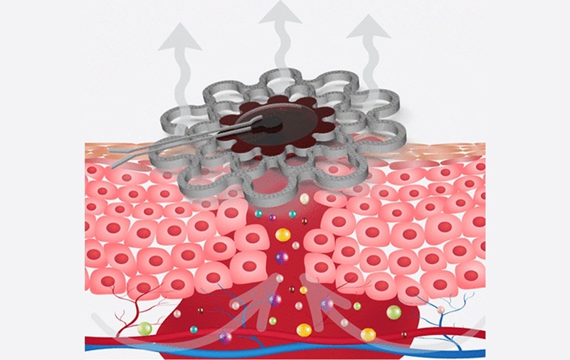
Specialized Dressing with Sensor Monitors pH Levels in Chronic Wounds
Any wound has the potential to become chronic, but the risk is significantly higher in individuals with certain medical conditions. Once a wound becomes chronic, healing slows, complications increase,... Read more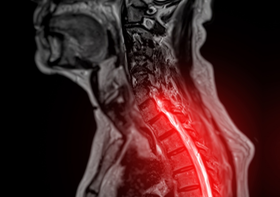
AI Model Could Help Diagnose Spinal Cord Disease Up To 30 Months Earlier
Cervical spondylotic myelopathy (CSM) is the leading cause of spinal cord dysfunction in older adults and occurs when arthritis in the neck compresses the spinal cord. The condition is chronic and progressive,... Read moreSurgical Techniques
view channel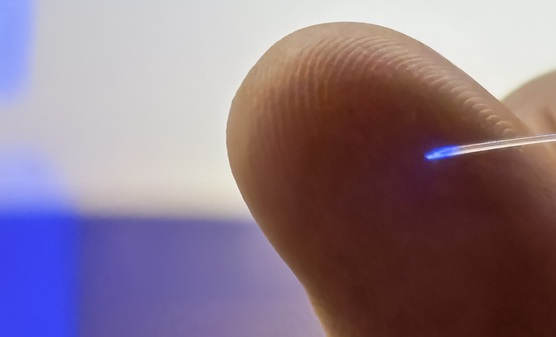
Brain Implant Records Neural Signals and Delivers Precise Medication
Neurological diseases such as epilepsy involve complex interactions across multiple layers of the brain, yet current implants can typically stimulate or record activity from only a single point.... Read moreAI-Based OCT Image Analysis Identifies High-Risk Plaques in Coronary Arteries
Lipid-rich plaques inside coronary arteries are strongly associated with heart attacks and other major cardiac events. While optical coherence tomography (OCT) provides detailed images of vessel structure... Read moreNeural Device Regrows Surrounding Skull After Brain Implantation
Placing electronic implants on the brain typically requires removing a portion of the skull, creating challenges for long-term access and safe closure. Current methods often involve temporarily replacing the skull or securing metal plates, which can lead to complications such as skin erosion and additional surgeries.... Read morePatient Care
view channel
Revolutionary Automatic IV-Line Flushing Device to Enhance Infusion Care
More than 80% of in-hospital patients receive intravenous (IV) therapy. Every dose of IV medicine delivered in a small volume (<250 mL) infusion bag should be followed by subsequent flushing to ensure... Read more
VR Training Tool Combats Contamination of Portable Medical Equipment
Healthcare-associated infections (HAIs) impact one in every 31 patients, cause nearly 100,000 deaths each year, and cost USD 28.4 billion in direct medical expenses. Notably, up to 75% of these infections... Read more
Portable Biosensor Platform to Reduce Hospital-Acquired Infections
Approximately 4 million patients in the European Union acquire healthcare-associated infections (HAIs) or nosocomial infections each year, with around 37,000 deaths directly resulting from these infections,... Read moreFirst-Of-Its-Kind Portable Germicidal Light Technology Disinfects High-Touch Clinical Surfaces in Seconds
Reducing healthcare-acquired infections (HAIs) remains a pressing issue within global healthcare systems. In the United States alone, 1.7 million patients contract HAIs annually, leading to approximately... Read moreHealth IT
view channel
EMR-Based Tool Predicts Graft Failure After Kidney Transplant
Kidney transplantation offers patients with end-stage kidney disease longer survival and better quality of life than dialysis, yet graft failure remains a major challenge. Although a successful transplant... Read more
Printable Molecule-Selective Nanoparticles Enable Mass Production of Wearable Biosensors
The future of medicine is likely to focus on the personalization of healthcare—understanding exactly what an individual requires and delivering the appropriate combination of nutrients, metabolites, and... Read moreBusiness
view channel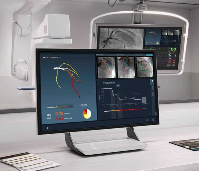
Medtronic to Acquire Coronary Artery Medtech Company CathWorks
Medtronic plc (Galway, Ireland) has announced that it will exercise its option to acquire CathWorks (Kfar Saba, Israel), a privately held medical device company, which aims to transform how coronary artery... Read more
Medtronic and Mindray Expand Strategic Partnership to Ambulatory Surgery Centers in the U.S.
Mindray North America and Medtronic have expanded their strategic partnership to bring integrated patient monitoring solutions to ambulatory surgery centers across the United States. The collaboration... Read more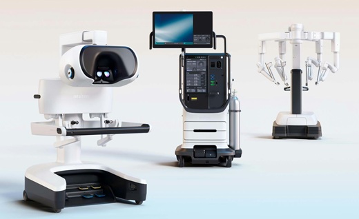
FDA Clearance Expands Robotic Options for Minimally Invasive Heart Surgery
Cardiovascular disease remains the world’s leading cause of death, with nearly 18 million fatalities each year, and more than two million patients undergo open-heart surgery annually, most involving sternotomy.... Read more