


Optical Virtual Biopsy Aids Robotic-Assisted Surgery
|
By HospiMedica International staff writers Posted on 22 Aug 2017 |
|
 system (Photo courtesy of Mauna Kea Technologies).")
Image: The Cellvizio probe-based confocal laser endomicroscopy (pCLE) system (Photo courtesy of Mauna Kea Technologies).
Ultra-High Definition (UHD) miniprobes provide visualization of body cavities, organs, and canals during endoscopic and laparoscopic surgical procedures, including robotic-assisted procedures.
The Mauna Kea Technologies (Suwanee, GA, USA) CelioFlex UHD confocal miniprobes are fiber optic probes designed to image the internal microstructure of tissues. CelioFlex UHD miniprobes are intended for use with the Cellvizio probe-based confocal laser endomicroscopy (pCLE) system in order to determine whether examined tissue is benign or malignant in patient management decisions. Embedded real-time image processing software combined with a high-speed laser scanning unit (LSU) allows the system to produce 12 frames per second (fps), so that the images stay sharp.
The system is designed so that only the flexible confocal miniprobes are inserted into the endoscope, leaving scanning and processing outside. The images thus come from a very thin focal plane that is optically projected into the tissues, a so-called “optical slicing” capability similar in principal to the physical slicing of endoscopically extracted tissues samples in standard histology. Probes are available for a wide range of conditions, including Barrett's Esophagus, Bilio-pancreatic strictures, pancreatic cysts, colorectal lesions, inflammatory bowel diseases, pulmonology and peripheral lung lesions.
“We see the ability to instantaneously visualize and characterize tissue at the cellular level as a natural extension to robotic-assisted procedures and enabling data-driven surgery, which will in-part rely on advanced imaging and machine learning algorithms to provide reliable, real-time tissue information to surgeons,” said Sacha Loiseau, PhD, founder and CEO of Mauna Kea Technologies.
pCLE is a diagnostic technique that allows microscopic examination of tissues during ongoing endoscopy. Different types of tissue and diseases can be diagnosed immediately, and analysis of the in vivo micro-architecture is helpful to better target standard biopsies and reduces the number of biopsies required. pCLE necessitates an intravenous injection of a fluorescent marker, such as fluorescein, to obtain a high level of magnification.
Related Links:
Mauna Kea Technologies
Gold Member
SARS‑CoV‑2/Flu A/Flu B/RSV Sample-To-Answer Test
SARS‑CoV‑2/Flu A/Flu B/RSV Cartridge (CE-IVD)
New
Medical Adhesive
MED 5570U
New
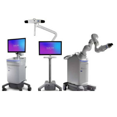
Surgical System
Stealth AXiS
Latest Surgical Techniques News
- Ultrasound Technology Aims to Replace Invasive BPH Procedures
- Continuous Monitoring with Wearables Enhances Postoperative Patient Safety
- New Approach Enables Customized Muscle Tissue Without Biomaterial Scaffolds
- Robot-Assisted Brain Angiography Improves Procedural Outcomes
- Brain Mapping Technology Enhances Precision in Brain Tumor Resection
- Handheld Robotic System Expands Options for Total Knee Surgery
- VR Experience Reduces Patient Anxiety Before Kidney Stone Procedure
- Injectable Mini Livers Offer Hope for Patients Awaiting Transplant
- Pulsed Field Ablation Technology Cleared in Europe for Persistent AFib
- AI-Powered Imaging Brings Real-Time Margin Clarity to Breast Cancer Surgery
- Minimally Invasive Device Safely Treats Challenging Brain Aneurysms
- Surgical Robot Makes Complex Liver Tumor Surgery Safer and Less Invasive
- Neurostimulation Implant Reduces Seizure Burden in Drug-Resistant Epilepsy
- Minimally Invasive Procedure Effectively Treats Small Kidney Cancers
- Fluorescence Probe Paired with Engineered Enzymes Lights Up Tumors for Easier Surgical Removal
- Novel Hydrogel Could Become Bone Implant of the Future
Channels
Artificial Intelligence
view channelAI Analysis of Pericardial Fat Refines Long-Term Heart Disease Risk
Accurately identifying long-term cardiovascular disease risk in asymptomatic adults remains challenging for clinicians. Missed or underestimated risk delays preventive therapy and increases the chance... Read more")
Machine Learning Approach Enhances Liver Cancer Risk Stratification
Hepatocellular carcinoma, the most common form of primary liver cancer, is often detected late despite targeted surveillance programs. Current screening guidelines emphasize patients with known cirrhosis,... Read more")
, k-means++ was used to identify clusters. Three clinically distinct clusters were identified. Among those clusters, patients were divided into solid organ transplant (SOT) and non-SOT groups. Clinical characteristics and outcomes were evaluated (Masayuki Nigo et al., American Journal of Transplantation (2026). DOI: 10.1016/j.ajt.2025.10.019)")
Critical Care
view channel")
Noninvasive Monitoring Device Enables Earlier Intervention in Heart Failure
Hospitalizations for heart failure with preserved ejection fraction (HFpEF) remain common because lung congestion often worsens before symptoms prompt treatment changes. Missed early decompensation... Read more")
Automated IV Labeling Solution Improves Infusion Safety and Efficiency
Medication administration in high-acuity settings is often complicated by multiple concurrent infusions, making accurate line identification essential. In a 10-hospital intensive care unit study, 60% of... Read more)")
")
Patient Care
view channel")
Wearable Sleep Data Predict Adherence to Pulmonary Rehabilitation
Chronic obstructive pulmonary disease (COPD) is a long-term lung disorder that makes breathing difficult and often disturbs sleep, reducing energy for daily activities. Limited engagement in pulmonary... Read more")
Revolutionary Automatic IV-Line Flushing Device to Enhance Infusion Care
More than 80% of in-hospital patients receive intravenous (IV) therapy. Every dose of IV medicine delivered in a small volume (<250 mL) infusion bag should be followed by subsequent flushing to ensure... Read more")
")
Health IT
view channel")
EMR-Based Tool Predicts Graft Failure After Kidney Transplant
Kidney transplantation offers patients with end-stage kidney disease longer survival and better quality of life than dialysis, yet graft failure remains a major challenge. Although a successful transplant... Read more")
Printable Molecule-Selective Nanoparticles Enable Mass Production of Wearable Biosensors
The future of medicine is likely to focus on the personalization of healthcare—understanding exactly what an individual requires and delivering the appropriate combination of nutrients, metabolites, and... Read more")
")
")
Business
view channel")
")
")