Researchers Develop AI Algorithm to Predict Immunotherapy Response
|
By HospiMedica International staff writers Posted on 12 Sep 2018 |
A team of French researchers have designed an algorithm and developed it to analyze Computed Tomography (CT) scan images, establishing for the first time that artificial intelligence (AI) can process medical images to extract biological and clinical information. The researchers have created a so-called radiomic signature, which defines the level of lymphocyte infiltration of a tumor and provides a predictive score for the efficacy of immunotherapy in the patient.
In the near future, this could make it possible for physicians to use imaging to identify biological phenomena in a tumor located anywhere in the body without performing a biopsy.
Currently, there are no markers, which can accurately identify patients who will respond to anti-PD-1/PD-L1 immunotherapy in a situation where only 15 to 30% of patients do respond to such treatment. The more immunologically richer the tumor environment (presence of lymphocytes), the higher is the chances of immunotherapy being effective. Hence, the researchers tried to characterize this environment using imaging and correlate this with the patients’ clinical response. In their study, the radiomic signature was captured, developed and validated genomically, histologically and clinically in 500 patients with solid tumors (all sites) from four independent cohorts.
The researchers first used a machine learning-based approach to teach the algorithm how to use relevant information extracted from CT scans of patients participating in an earlier study, which also held tumor genome data. Thus, based solely on images, the algorithm learned to predict what the genome might have revealed about the tumor immune infiltrate, in particular with respect to the presence of cytotoxic T-lymphocytes (CD8) in the tumor, thus establishing a radiomic signature.
The researchers tested and validated this signature in other cohorts, including that of TCGA (The Cancer Genome Atlas), thus demonstrating that imaging could predict a biological phenomenon, providing an estimation of the degree of immune infiltration of a tumor. Further, in order to test the signature’s applicability in a real situation and correlate it to the efficacy of immunotherapy, it was evaluated using CT scans performed before the start of treatment in patients participating in five phase I trials of anti-PD-1/PD-L1 immunotherapy. The researchers found that the patients in whom immunotherapy was effective at three and six months had higher radiomic scores as did those with better overall survival.
In their next clinical study, the researchers will assess the signature both retrospectively and prospectively, using a larger number of patients and stratifying them based on cancer type in order to refine the signature. They will also use more sophisticated automatic learning and AI algorithms to predict patient response to immunotherapy, while integrating data from imaging, molecular biology and tissue analysis. The researchers aim to identify those patients who are most likely to respond to treatment, thereby improving the efficacy/cost ratio of treatment.
Channels
Artificial Intelligence
view channelCritical Care
view channel
New Inhalable Treatment for TB Lowers Side Effects
Tuberculosis (TB) remains one of the world’s deadliest infectious diseases, despite being curable. Treatment requires months of multiple drugs that can cause serious side effects, leading many patients... Read more
AI Algorithm Improves Antibiotic Decision-Making in Urinary Tract Infection
Urinary tract infections (UTIs) are among the most common bacterial infections worldwide and are a major driver of antibiotic use. Inappropriate or overly broad prescribing contributes to antimicrobial... Read more
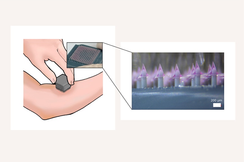
3D-Printed System Enhances Vaccine Delivery Via Microneedle Array Patch
The COVID-19 pandemic underscored the need for efficient, durable, and widely accessible vaccines. Conventional vaccination requires trained personnel and cold-chain logistics, which can slow mass immunization... Read more
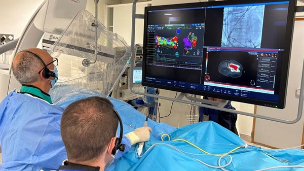
Whole-Heart Mapping Technology Provides Comprehensive Real-Time View of Arrhythmias
Cardiac arrhythmias can be difficult to diagnose and treat because current mapping systems analyze the heart one chamber at a time. This fragmented view forces clinicians to infer electrical activity they... Read moreSurgical Techniques
view channelAI-Based OCT Image Analysis Identifies High-Risk Plaques in Coronary Arteries
Lipid-rich plaques inside coronary arteries are strongly associated with heart attacks and other major cardiac events. While optical coherence tomography (OCT) provides detailed images of vessel structure... Read moreNeural Device Regrows Surrounding Skull After Brain Implantation
Placing electronic implants on the brain typically requires removing a portion of the skull, creating challenges for long-term access and safe closure. Current methods often involve temporarily replacing the skull or securing metal plates, which can lead to complications such as skin erosion and additional surgeries.... Read morePatient Care
view channel
Revolutionary Automatic IV-Line Flushing Device to Enhance Infusion Care
More than 80% of in-hospital patients receive intravenous (IV) therapy. Every dose of IV medicine delivered in a small volume (<250 mL) infusion bag should be followed by subsequent flushing to ensure... Read more
VR Training Tool Combats Contamination of Portable Medical Equipment
Healthcare-associated infections (HAIs) impact one in every 31 patients, cause nearly 100,000 deaths each year, and cost USD 28.4 billion in direct medical expenses. Notably, up to 75% of these infections... Read more
Portable Biosensor Platform to Reduce Hospital-Acquired Infections
Approximately 4 million patients in the European Union acquire healthcare-associated infections (HAIs) or nosocomial infections each year, with around 37,000 deaths directly resulting from these infections,... Read moreFirst-Of-Its-Kind Portable Germicidal Light Technology Disinfects High-Touch Clinical Surfaces in Seconds
Reducing healthcare-acquired infections (HAIs) remains a pressing issue within global healthcare systems. In the United States alone, 1.7 million patients contract HAIs annually, leading to approximately... Read moreHealth IT
view channel
EMR-Based Tool Predicts Graft Failure After Kidney Transplant
Kidney transplantation offers patients with end-stage kidney disease longer survival and better quality of life than dialysis, yet graft failure remains a major challenge. Although a successful transplant... Read more
Printable Molecule-Selective Nanoparticles Enable Mass Production of Wearable Biosensors
The future of medicine is likely to focus on the personalization of healthcare—understanding exactly what an individual requires and delivering the appropriate combination of nutrients, metabolites, and... Read moreBusiness
view channel
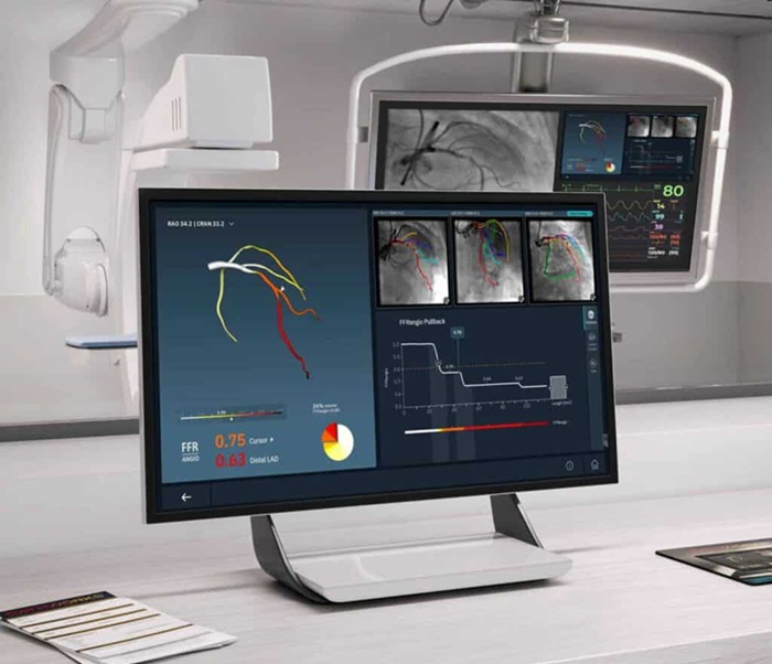
Medtronic to Acquire Coronary Artery Medtech Company CathWorks
Medtronic plc (Galway, Ireland) has announced that it will exercise its option to acquire CathWorks (Kfar Saba, Israel), a privately held medical device company, which aims to transform how coronary artery... Read more
Medtronic and Mindray Expand Strategic Partnership to Ambulatory Surgery Centers in the U.S.
Mindray North America and Medtronic have expanded their strategic partnership to bring integrated patient monitoring solutions to ambulatory surgery centers across the United States. The collaboration... Read more
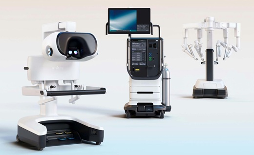
FDA Clearance Expands Robotic Options for Minimally Invasive Heart Surgery
Cardiovascular disease remains the world’s leading cause of death, with nearly 18 million fatalities each year, and more than two million patients undergo open-heart surgery annually, most involving sternotomy.... Read more


