



Rerouting Nerves During Amputation Reduces Phantom Limb Pain
|
By HospiMedica International staff writers Posted on 09 Jan 2019 |


|
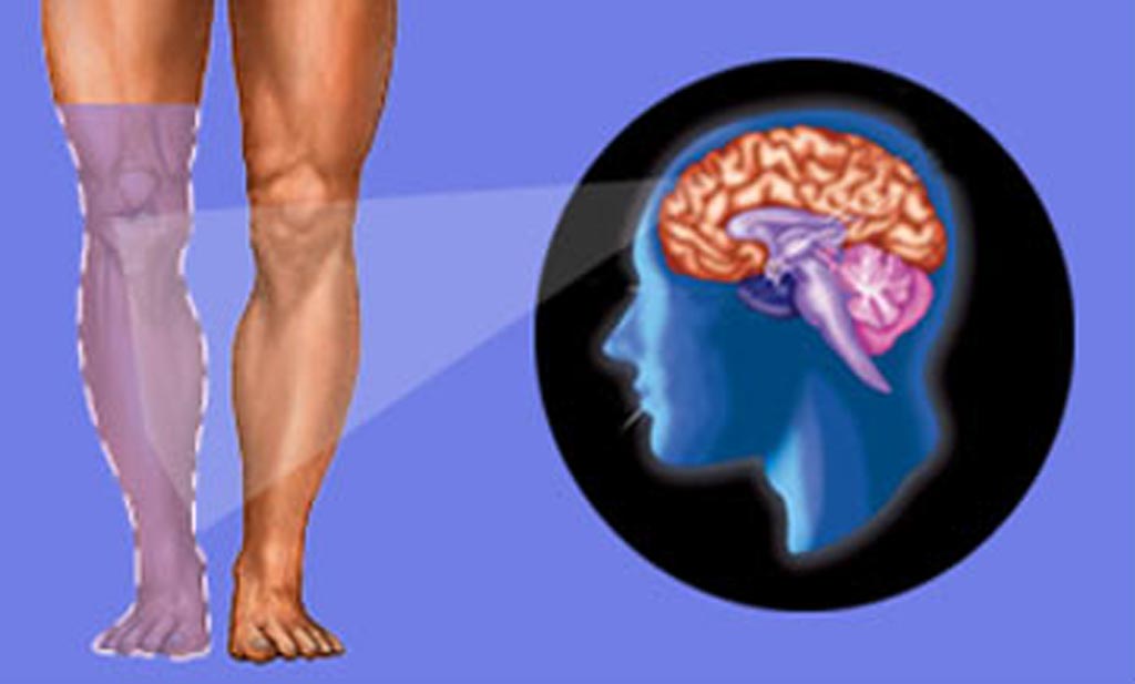
Image: A new study asserts that redirecting nerves severed during limb amputation can reduce phantom pain (Photo courtesy of 123rf).
A novel surgical procedure developed for advanced prosthetics can prevent or reduce debilitating phantom limb and stump pain in amputees, according to a new study.
Researchers at Ohio State University (OSU; Columbus, USA) conducted a study in 22 patients in order to evaluate the results of primary targeted muscle reinnervation (TMR) for below-the-knee amputations. The procedure involves rerouting the severed motor nerves by surgically transferring them to the motor points of the denervated target muscles, which, after reinnervation, can contract in response to neural control signals intended for the missing limb. The operation of the prosthesis thus occurs in response to attempts to move the missing limb, making control easier and more intuitive.
All subjects denied neuroma pain following amputation. Each patient was followed on an outpatient basis for one year to evaluate early symptoms of neuroma or phantom limb pain, patient satisfaction, and functionality. The researchers also found that over the course of three years, none of the patients developed symptomatic neuromas, and only 13% of those who received primary TMR reported pain six months later. In addition, by creating the additional control sites, TMR eliminated the need to switch the prosthesis between different control modes. The study was published in the January 2019 issue of Plastic and Reconstructive Surgery.
“A significant amount of pain in amputees is caused by disorganized nerve endings, i.e. symptomatic neuromas, in the residual limb. They form when nerves are severed and not addressed, thus they have nowhere to go,” said senior author Ian Valerio, MD, of the department of plastic and reconstructive surgery at OSU. “Attaching those cut nerve endings to motor nerves in a nearby muscle allows the body to re-establish its neural circuitry. This alleviates phantom and residual limb pain by giving those severed nerves somewhere to go and something to do.”
“TMR allows for more individual muscle unit firings through the patient's thoughts. It provides for better intuitive control, resulting in more refined functional movements and more degrees of motion by an advanced prosthetic,” concluded lead author J. Byers Bowen, MD, of OSU. “When done at the time of initial amputation, there is minimal health risk, and recovery is similar to that of traditional amputation surgery.”
Approximately 25% of major limb amputees will develop chronic localized symptomatic neuromas and phantom limb pain in the residual stump or limb. Previous studies show that secondary targeted reinnervation successfully treats and, in some cases, resolves peripheral neuropathy and phantom limb pain in patients who have undergone a previous amputation.
Related Links:
Ohio State University





Researchers at Ohio State University (OSU; Columbus, USA) conducted a study in 22 patients in order to evaluate the results of primary targeted muscle reinnervation (TMR) for below-the-knee amputations. The procedure involves rerouting the severed motor nerves by surgically transferring them to the motor points of the denervated target muscles, which, after reinnervation, can contract in response to neural control signals intended for the missing limb. The operation of the prosthesis thus occurs in response to attempts to move the missing limb, making control easier and more intuitive.
All subjects denied neuroma pain following amputation. Each patient was followed on an outpatient basis for one year to evaluate early symptoms of neuroma or phantom limb pain, patient satisfaction, and functionality. The researchers also found that over the course of three years, none of the patients developed symptomatic neuromas, and only 13% of those who received primary TMR reported pain six months later. In addition, by creating the additional control sites, TMR eliminated the need to switch the prosthesis between different control modes. The study was published in the January 2019 issue of Plastic and Reconstructive Surgery.
“A significant amount of pain in amputees is caused by disorganized nerve endings, i.e. symptomatic neuromas, in the residual limb. They form when nerves are severed and not addressed, thus they have nowhere to go,” said senior author Ian Valerio, MD, of the department of plastic and reconstructive surgery at OSU. “Attaching those cut nerve endings to motor nerves in a nearby muscle allows the body to re-establish its neural circuitry. This alleviates phantom and residual limb pain by giving those severed nerves somewhere to go and something to do.”
“TMR allows for more individual muscle unit firings through the patient's thoughts. It provides for better intuitive control, resulting in more refined functional movements and more degrees of motion by an advanced prosthetic,” concluded lead author J. Byers Bowen, MD, of OSU. “When done at the time of initial amputation, there is minimal health risk, and recovery is similar to that of traditional amputation surgery.”
Approximately 25% of major limb amputees will develop chronic localized symptomatic neuromas and phantom limb pain in the residual stump or limb. Previous studies show that secondary targeted reinnervation successfully treats and, in some cases, resolves peripheral neuropathy and phantom limb pain in patients who have undergone a previous amputation.
Related Links:
Ohio State University

Gold Member
STI Test
Vivalytic Sexually Transmitted Infection (STI) Array
Hybrid Arch Device
Neo EDE
Radiofrequency Generator
GX1
Latest Surgical Techniques News
- New Ultrasonic Surgical Platform Adds Control for Bone and Soft Tissue Removal
- Targeted Radiotherapy Before Surgery Improves Control of Kidney Cancer Thrombus
- New Surgical Technique Enlarges Aortic Root for Larger Valve Implantation

- FDA Approval Expands Bridging Stent Use in Complex Aortic Aneurysm Repair
- New Beam Measurement Approach Could Support More Precise Ion Therapy
- Tiny Robots Could Speed Targeted Drug Delivery to Heart Infections
- Contact Radiotherapy Offers New Organ-Preserving Approach for Rectal Cancer
- FDA Clears Antibiotic-Delivering Spacer for Infected Knee Replacements
- Vascular Occlusion System Gains Expanded FDA Indication for Venous Embolization
- Covered Stent System Achieves High Nine-Month Patency in Iliac Artery Disease
- New Heart Recovery Approach May Broaden Pediatric Transplant Donor Pool
- Matrix-Based Skin Substitute Improves Closure of Diabetic Foot Ulcers
- Injectable Foam Dressing Conforms to Deep Traumatic Wounds and Inhibits Bacteria
- Magnetically Guided Capsule Enables Targeted Small Intestine Microbiome Sampling
- New FDA-Approved Pediatric Pulmonary Valve Can Expand as Children Grow
- New Software Tools Support Precision Mapping and Guidance in Cardiac Ablation
Channels
Artificial Intelligence
view channel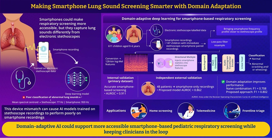
AI Tool Improves Smartphone Detection of Abnormal Pediatric Lung Sounds
Respiratory disease in children remains a major clinical concern worldwide. Abnormal lung sounds can indicate pediatric respiratory illness, but standard auscultation usually requires an in-person examination... Read more
AI Tool Detects Coronary Heart Disease from Face and Tongue Images
Coronary heart disease (CHD) develops when fatty plaques narrow the arteries supplying the heart muscle, restricting blood flow and sometimes first presenting as chest pain, myocardial infarction, or another... Read more

Critical Care
view channel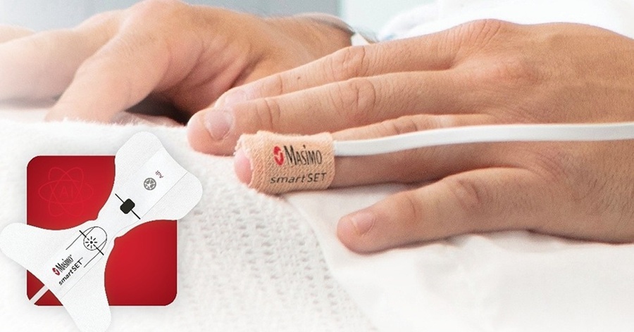
Smart Pulse Oximetry Supports Earlier Recognition of Arrhythmias and Respiratory Risk
On general care floors, respiratory compromise and arrhythmias can develop between routine checks. Atrial fibrillation often goes undiagnosed in older adults, while opioid-induced respiratory depression... Read more
Proposed Risk Score Prioritizes Sickest Patients for Heart Transplant
Donor hearts remain extremely scarce in the U.S. transplant system, with one in five candidates dying or being removed from the waiting list before receiving an organ. Current allocation rules may also... Read more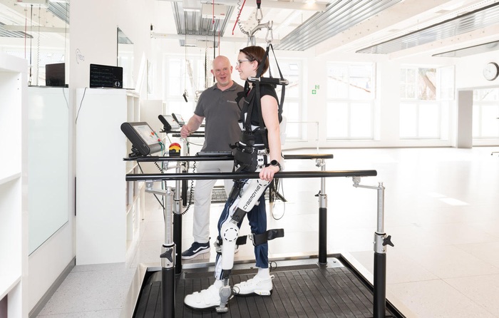

Patient Care
view channel
AI Platform Personalizes Support Between Visits for Cancer Survivors
Cancer survivors often manage persistent side effects after treatment, including fatigue, anxiety, uncertainty, symptoms, stress, and questions between appointments. Clinical teams need ways to maintain... Read more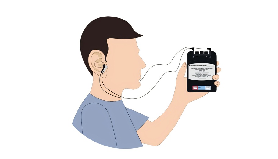
At-Home Neuromodulation Shows Promise in Selected Patients With Hypertension
High blood pressure is a major risk factor for cardiovascular disease, including heart attack and stroke. Many patients receiving treatment for hypertension still remain above recommended blood pressure... Read more
EHR Model Flags High-Risk Periods in Patients with Metastatic Breast Cancer
Determining when patients with metastatic breast cancer are nearing the end of life remains difficult, often leading to intensive interventions with limited benefit. Prognostic uncertainty can delay goals-of-care... Read more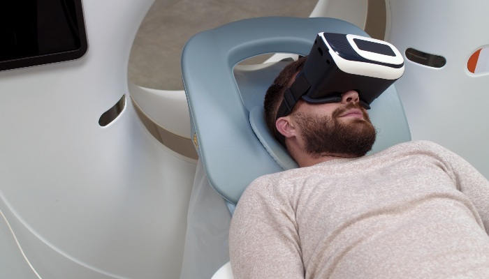
Medical Imaging
view channel
Amyloid and Tau PET Guide Selection and Monitoring of Alzheimer’s Therapy
Alzheimer’s disease is the most common form of dementia and can be difficult to distinguish from other causes of cognitive decline. According to the World Health Organization (WHO), 57 million people were... Read more
MRI Tool Analysis Identifies Vascular Differences in Cerebral Small Vessel Disease
Cerebral small vessel disease (CSVD) involves changes or damage to the brain’s small blood vessels and is associated with stroke and cognitive impairment. Magnetic resonance imaging (MRI) can show cerebral... Read more
Health IT
view channel
EHR Alerts Support Dosing, Sedation, and Shock Recognition in Cardiac Care
Electronic health record alerts can add to clinician workload when they are poorly targeted. In critical cardiac care, however, delayed decisions can affect treatment timing, sedation exposure, and escalation... Read more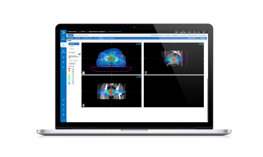
Adaptive Radiation QA Software Gains Validation for NeoArc Treatments
Adaptive radiation therapy allows clinicians to adjust treatment plans to daily anatomical changes such as tumor shrinkage, weight loss, or organ movement, improving precision while limiting exposure to... Read more

Point of Care
view channel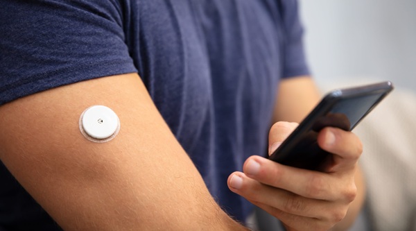
Continuous Glucose Monitoring Identifies Cardiometabolic Risk in Adults Without Diabetes
Dysglycemia—abnormal blood glucose regulation—can fluctuate throughout the day and often escape conventional screening. Clinicians typically rely on fasting plasma glucose and hemoglobin A1c, which offer... Read more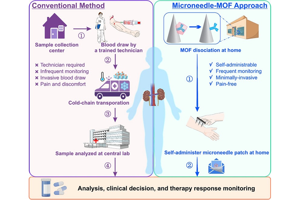
Microneedle Patch Enables At-Home Monitoring of Acute Kidney Injury
Kidney disease is often silent in its early stages, making timely detection difficult outside clinical settings. Early biomarkers such as neutrophil gelatinase–associated lipocalin can require blood draws... Read more
Business
view channel
Steris to Expand Surgical Safety Portfolio With $2.1 Billion Xodus Medical Deal
Steris has signed a definitive agreement to acquire Xodus Medical, a specialty surgical products business based in Pennsylvania, for $2.1 billion at closing. The acquisition will expand Steris’s healthcare... Read more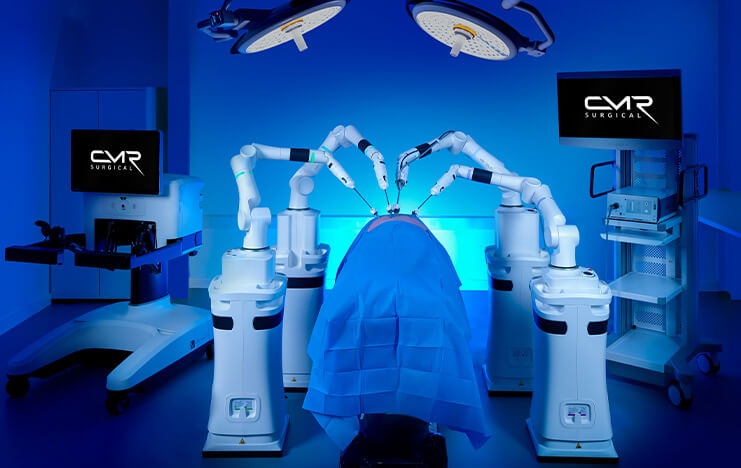
Stryker Enters Soft-Tissue Robotics Through Strategic Alliance With CMR Surgical
Stryker (Portage, MI, USA) and CMR Surgical (Cambridge, UK) have announced a strategic alliance centered on the Versius Surgical Robotic portfolio. The agreement includes an approximately USD 400 million... Read more
Terumo Neuro Acquisition Advances Liquid Embolic Technology for Chronic Subdural Hematoma
Terumo Neuro (Aliso Viejo, CA, USA) signed an agreement to acquire Arsenal Medical Inc. (Waltham, MA, USA), a privately held, clinical-stage biomaterials company. The proposed acquisition would add NeoCast,... Read more