New AI System Can Diagnose and Classify Intracranial Hemorrhage
|
By HospiMedica International staff writers Posted on 23 Jan 2019 |
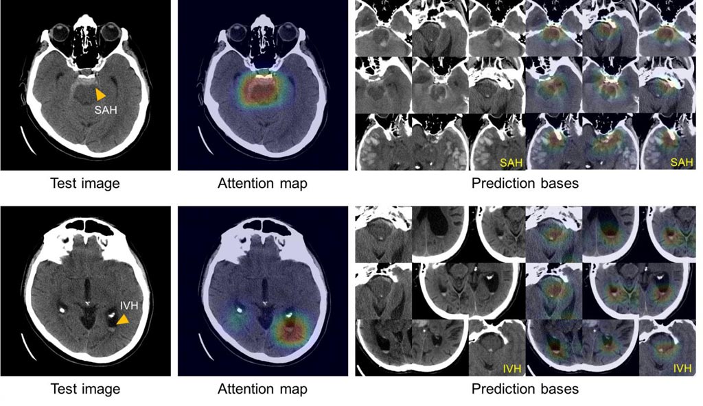
Image: These images show the system’s ability to explain its diagnosis of subarachnoid (left above) and intraventricular (left below) hemorrhage by displaying images with similar appearances (right) from an atlas of images used to train the system (Photo courtesy of Hyunkwang Lee, Harvard School of Engineering and Applied Sciences, and Sehyo Yune, MD, Massachusetts General Hospital Department of Radiology).
Researchers from the Massachusetts General Hospital (Boston, MA, USA) have developed an artificial intelligence (AI) system that can quickly diagnose and classify brain hemorrhages, as well as provide the basis of its decisions from relatively small image datasets. The system could help hospital emergency departments in evaluating patients with symptoms of a potentially life-threatening stroke, allowing rapid application of the correct treatment.
The researchers trained the system with 904 head CT scans, each consisting of around 40 individual images, that were labeled by neuroradiologists as to whether they depicted one of five hemorrhage subtypes, based on the location within the brain, or no hemorrhage. The team improved the accuracy of the deep-learning system by building in steps mimicking the way radiologists analyze images. These included adjusting factors such as contrast and brightness to reveal subtle differences not immediately apparent and scrolling through adjacent CT scan slices to determine whether or not something that appears on a single image reflects a real problem or is a meaningless artifact.
After the model system was created, the investigators tested it on two separate sets of CT scans – a retrospective set taken before the system was developed, consisting of 100 scans with and 100 without intracranial hemorrhage, and a prospective set of 79 scans with and 117 without hemorrhage, taken after the model was created. In its analysis of the retrospective set, the model system was as accurate in detecting and classifying intracranial hemorrhages as the radiologists that had reviewed the scans had been. In its analysis of the prospective set, the system proved to be even better than non-expert human readers.
To solve the “black box” problem, or the inability of systems to explain how they arrived at a decision, the team had the system review and save the images from the training dataset that most clearly represented the classic features of each of the five hemorrhage subtypes. Using this atlas of distinguishing features, the system is able to display a group of images similar to those of the CT scan being analyzed in order to explain the basis of its decisions.
“Rapid recognition of intracranial hemorrhage, leading to prompt appropriate treatment of patients with acute stroke symptoms, can prevent or mitigate major disability or death,” said co-author Michael Lev, MD, MGH Radiology. “Many facilities do not have access to specially trained neuroradiologists – especially at night or over weekends – which can require non-expert providers to determine whether or not a hemorrhage is the cause of a patient’s symptoms. The availability of a reliable, ‘virtual second opinion’ – trained by neuroradiologists – could make those providers more efficient and confident and help ensure that patients get the right treatment.”
“In addition to providing that much needed virtual second opinion, this system also could be deployed directly onto scanners, alerting the care team to the presence of a hemorrhage and triggering appropriate further testing before the patient is even off the scanner,” added co-author Shahein Tajmir, MD, MGH Radiology. “The next step will be to deploy the system into clinical areas and further validate its performance with many more cases. We are currently building a platform to allow for the widespread application of such tools throughout the department. Once we have this running in the clinical setting, we can evaluate its impact on turnaround time, clinical accuracy and the time to diagnosis.”
Related Links:
Massachusetts General Hospital
Latest Industry News News
Channels
Critical Care
view channel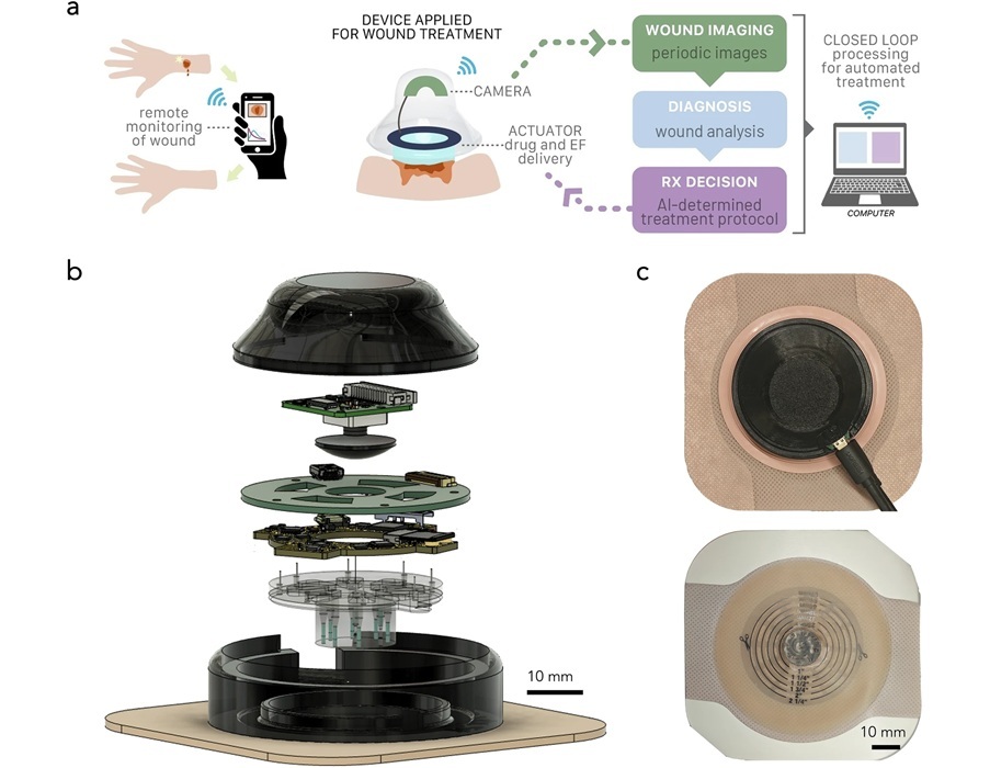
Wearable ‘Microscope in a Bandage’ Fastens Wound Healing
Wound healing is a complex biological process that moves through stages, including clotting, immune response, scabbing, and scarring. For many patients, especially those in remote areas or with limited... Read more
Virus Cocktail to Combat Superbugs Offers New Precision Medicine Approach for Hospitals Battling AMR
Antimicrobial resistance is one of the most pressing challenges in modern medicine, making once-treatable infections increasingly lethal. Enterobacter infections, for example, are difficult to treat and... Read moreSurgical Techniques
view channel
3D Printable Bio-Active Glass Could Serve as Bone Replacement Material
Glass may not seem like a natural choice for replacing bone, yet the two materials share surprising similarities in structure and strength. Bone and glass both bear weight more effectively than they withstand... Read more
Micro Imaging Device Paired with Endoscope Spots Cancers at Earlier Stage
Digestive system cancers are among the most common cancers, with hundreds of thousands of new cases and deaths reported annually in the United States. Standard endoscopy, the main diagnostic method for... Read more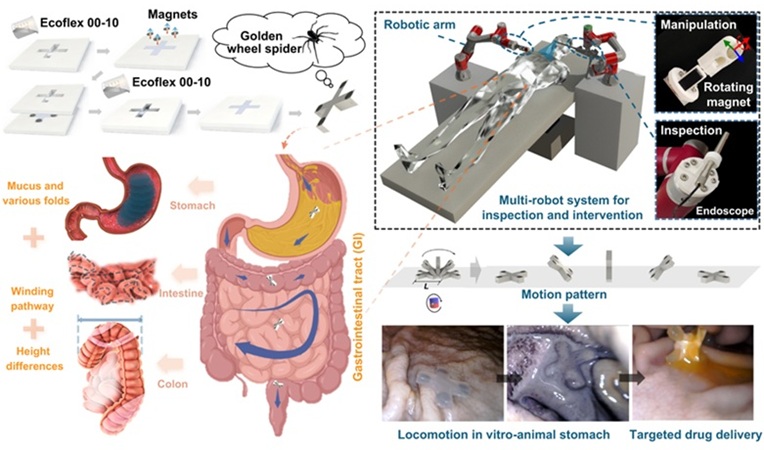
Spider-Inspired Magnetic Soft Robots to Perform Minimally Invasive GI Tract Procedures
The gastrointestinal (GI) tract is vital for digestion, nutrient absorption, and waste elimination, but it is also prone to cancers and other serious conditions. Standard endoscopy is widely used for diagnosis... Read morePatient Care
view channel
Revolutionary Automatic IV-Line Flushing Device to Enhance Infusion Care
More than 80% of in-hospital patients receive intravenous (IV) therapy. Every dose of IV medicine delivered in a small volume (<250 mL) infusion bag should be followed by subsequent flushing to ensure... Read more
VR Training Tool Combats Contamination of Portable Medical Equipment
Healthcare-associated infections (HAIs) impact one in every 31 patients, cause nearly 100,000 deaths each year, and cost USD 28.4 billion in direct medical expenses. Notably, up to 75% of these infections... Read more
Portable Biosensor Platform to Reduce Hospital-Acquired Infections
Approximately 4 million patients in the European Union acquire healthcare-associated infections (HAIs) or nosocomial infections each year, with around 37,000 deaths directly resulting from these infections,... Read moreFirst-Of-Its-Kind Portable Germicidal Light Technology Disinfects High-Touch Clinical Surfaces in Seconds
Reducing healthcare-acquired infections (HAIs) remains a pressing issue within global healthcare systems. In the United States alone, 1.7 million patients contract HAIs annually, leading to approximately... Read moreHealth IT
view channel
Printable Molecule-Selective Nanoparticles Enable Mass Production of Wearable Biosensors
The future of medicine is likely to focus on the personalization of healthcare—understanding exactly what an individual requires and delivering the appropriate combination of nutrients, metabolites, and... Read moreBusiness
view channel
Philips and Masimo Partner to Advance Patient Monitoring Measurement Technologies
Royal Philips (Amsterdam, Netherlands) and Masimo (Irvine, California, USA) have renewed their multi-year strategic collaboration, combining Philips’ expertise in patient monitoring with Masimo’s noninvasive... Read more
B. Braun Acquires Digital Microsurgery Company True Digital Surgery
The high-end microsurgery market in neurosurgery, spine, and ENT is undergoing a significant transformation. Traditional analog microscopes are giving way to digital exoscopes, which provide improved visualization,... Read more
CMEF 2025 to Promote Holistic and High-Quality Development of Medical and Health Industry
The 92nd China International Medical Equipment Fair (CMEF 2025) Autumn Exhibition is scheduled to be held from September 26 to 29 at the China Import and Export Fair Complex (Canton Fair Complex) in Guangzhou.... Read more


