


AI Software Diagnoses Lung Cancer from CT Images
|
By HospiMedica International staff writers Posted on 07 Mar 2019 |
|
.")
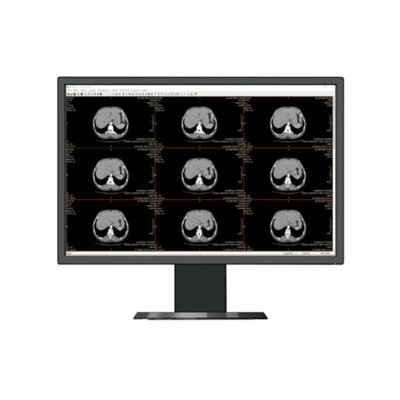
Image: The Doctor AIzimov system analyzes computed tomography results within 20 seconds (Photo courtesy of Peter the Great Saint-Petersburg Polytechnic University).
A team of Russian researchers, in collaboration with radiologists, has developed an intelligent software system for lung cancer diagnostics. The software, which can be installed on any computer, analyzes patients' computed tomography (CT) results within 20 seconds and provides an image in which the pathology is clearly marked. The system has been named Doctor AIzimov (AI for Artificial Intelligence) in honor of the science-fiction writer Isaac Asimov, who developed three famous laws of robotics.
The first tests of this intelligent system were carried out at the end of 2018 in which the system analyzed CT images of 60 patients and found focal nodules in lungs of small sizes (2 mm). The open testing of the intelligent system will be carried out at the beginning of 2019. The system will be adapted to analyze the results of the ultrasound and X-ray medical investigation of other organs.
In a new proposed and developed approach to the lung cancer classification using the chord method, segmented CT images are used: points are randomly drawn on the surface of a nodule, after that the points are connected by lines (chords). The length histogram of the chords reflects the shape and structure of the tumor. Although the system examines every nodule from the inside, its external surroundings are also very important. To learn more about the tumor, it is placed in a cube, and perpendiculars are drawn from its edges to the surface of the nodule.
Thus, instead of classifying graphically complex and heavy images of the CT (the size of every CT image is approximately 1 GB), the nodule is represented in the form of compact and simple histograms, which are then analyzed by the Doctor AIzimov system. The scientists have also trained the system to distinguish malignant and benign tumors. Currently, the dataset holds CT images of about 250 patients and the scientists plan to increase the number of images by four times by mid-2019.
With each new CT image, the system self-improves. In the future, a patient's CT images will be transferred to the supercomputer using the Internet that will reduce the diagnostic testing time per patient from 20 till 2 seconds. After that a radiologist will receive the marked image instead of the large CT image, thus significantly reducing the time needed for the analysis and diagnostics.
“Many different objects may be detected on the CT images, so the main task was to train the system to recognize what each of the objects represents. Using the clinical and radiological classification, we are trying to train the system not only to detect tumors, but also to distinguish other diseases similar to cancer,” said Anna Meldo, the head of the Radiology Department of the St. Petersburg Clinical Research Center for Specialized Types of Medical Care (Oncological).
Gold Member
SARS‑CoV‑2/Flu A/Flu B/RSV Sample-To-Answer Test
SARS‑CoV‑2/Flu A/Flu B/RSV Cartridge (CE-IVD)
New
Medical-Grade Display
HL2316SHTB
New
Multi-Chamber Washer-Disinfector
WD 390
Latest Industry News News
Channels
Artificial Intelligence
view channelAI Analysis of Pericardial Fat Refines Long-Term Heart Disease Risk
Accurately identifying long-term cardiovascular disease risk in asymptomatic adults remains challenging for clinicians. Missed or underestimated risk delays preventive therapy and increases the chance... Read more")
Machine Learning Approach Enhances Liver Cancer Risk Stratification
Hepatocellular carcinoma, the most common form of primary liver cancer, is often detected late despite targeted surveillance programs. Current screening guidelines emphasize patients with known cirrhosis,... Read more")
, k-means++ was used to identify clusters. Three clinically distinct clusters were identified. Among those clusters, patients were divided into solid organ transplant (SOT) and non-SOT groups. Clinical characteristics and outcomes were evaluated (Masayuki Nigo et al., American Journal of Transplantation (2026). DOI: 10.1016/j.ajt.2025.10.019)")
Critical Care
view channel")
Angiography-Based FFR Approach Matches Gold Standard Results Without Wires
Accurately determining whether a coronary stenosis limits blood flow is essential to guide percutaneous coronary intervention, yet wire-based physiologic testing remains underused due to added procedural... Read more")
Eye Imaging AI Identifies Elevated Cardiovascular Risk
Many adults at risk for atherosclerotic cardiovascular disease are not identified until they undergo formal primary care assessment. Delayed risk recognition can postpone initiation of statins and lifestyle... Read more")
")
Surgical Techniques
view channel")
Fiber-Form Bone Graft Expands Intraoperative Options for Spinal Fusion
Spinal and orthopedic fusion procedures often require bone graft materials that handle predictably and support bone formation. Surgeons face added complexity in difficult anatomy and challenging fusion environments.... Read more")
Ultrasound‑Aided Catheter Treatment Cuts Early Collapse in Pulmonary Embolism
Acute pulmonary embolism can cause rapid hemodynamic deterioration and early death in hospitalized and emergency patients. Systemic thrombolysis can dissolve clots but is limited by a high risk of major... Read more")
")
Patient Care
view channel")
Wearable Sleep Data Predict Adherence to Pulmonary Rehabilitation
Chronic obstructive pulmonary disease (COPD) is a long-term lung disorder that makes breathing difficult and often disturbs sleep, reducing energy for daily activities. Limited engagement in pulmonary... Read more")
Revolutionary Automatic IV-Line Flushing Device to Enhance Infusion Care
More than 80% of in-hospital patients receive intravenous (IV) therapy. Every dose of IV medicine delivered in a small volume (<250 mL) infusion bag should be followed by subsequent flushing to ensure... Read more")
")
Health IT
view channel")
Voice-Driven AI System Enables Structured GI Procedure Documentation
Documentation during gastrointestinal (GI) procedures often competes with real-time clinical decision-making and imposes a significant cognitive burden on physicians. Manual data entry and post-procedure... Read more")
EMR-Based Tool Predicts Graft Failure After Kidney Transplant
Kidney transplantation offers patients with end-stage kidney disease longer survival and better quality of life than dialysis, yet graft failure remains a major challenge. Although a successful transplant... Read more")
Printable Molecule-Selective Nanoparticles Enable Mass Production of Wearable Biosensors
The future of medicine is likely to focus on the personalization of healthcare—understanding exactly what an individual requires and delivering the appropriate combination of nutrients, metabolites, and... Read more")
")
Business
view channel")
")
")