


Skin Bioprinting System Helps Heal Extensive Wounds
|
By HospiMedica International staff writers Posted on 12 Mar 2019 |


|
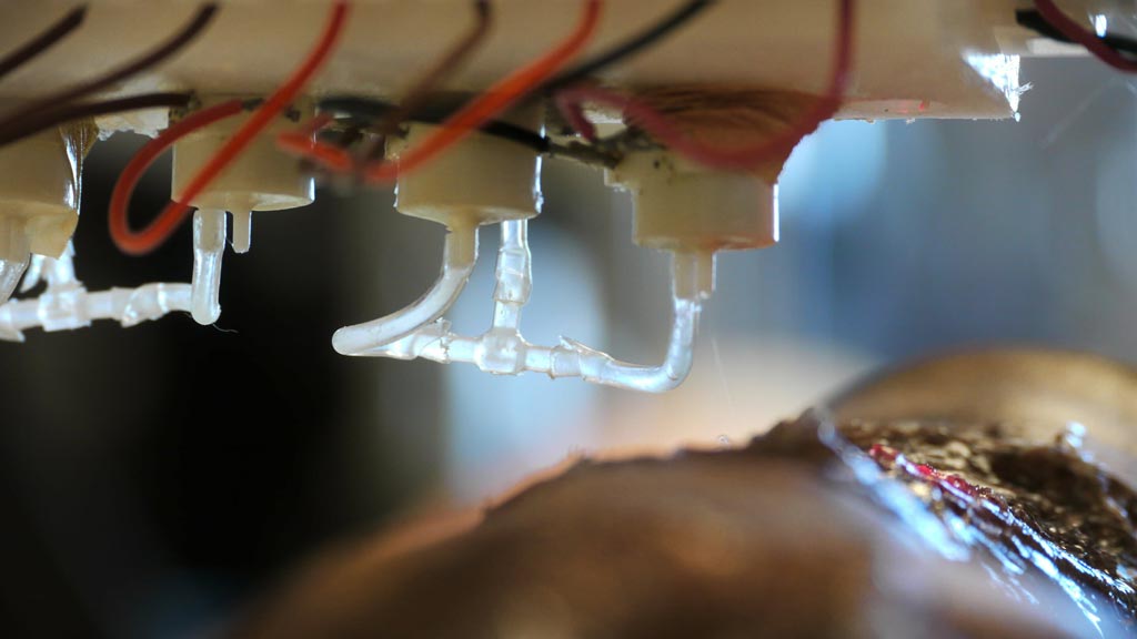
Image: A close up of the nozzle of the skin bioprinter (Photo courtesy of WFIRM).
A mobile skin bioprinter that layers dermal fibroblasts and epidermal keratinocytes directly into a wound can accelerate contraction and re-epithelialization, claims a new study.
Developed at the Wake Forest Institute for Regenerative Medicine (WFIRM; Winston-Salem, NC, USA), the mobile bioprinting system first isolates the major skin cells--dermal fibroblasts and epidermal keratinocytes-- from a small biopsy of uninjured tissue. The cells are then expanded and mixed into a hydrogel and placed into the bioprinter. Using integrated imaging technology the wound is then scanned; the data is used to direct the print heads on which cells to deliver exactly where in the wound, layer by layer.
The process thus replicates and accelerates the formation of normal skin structure and function. The fibroblasts synthesize extracellular matrix (ECM) and collagen, which play a critical role in wound healing, while the keratinocytes reconstruct the epidermis, the outermost layer of the skin. The regenerated tissues show a dermal structure and composition similar to healthy skin, with extensive collagen deposition arranged in large, organized fibers, extensive mature vascular formation, and proliferating keratinocytes. The study was published on February 12, 2019, in Nature Scientific Reports.
“The unique aspect of this technology is the mobility of the system and the ability to provide on-site management of extensive wounds by scanning and measuring them in order to deposit the cells directly where they are needed to create skin,” said lead author Sean Murphy, PhD. “Currently, skin grafts to treat wounds and burns are the ‘gold standard’ technique, but adequate coverage of wounds is often a challenge, particularly when there is limited availability of healthy skin to harvest.”
The early treatment and rapid closure of acute or chronic wounds is essential for normal healing and prevention of hypertrophic scarring; but the use of split thickness autografts is often limited by the availability of a suitable area of healthy donor skin to harvest. Cellular and non-cellular biological skin-equivalents are commonly used as an alternative treatment option for these patients, however these treatments usually involve multiple surgical procedures and are associated with high costs of production and repeated wound treatment.
Related Links:
Wake Forest Institute for Regenerative Medicine






Developed at the Wake Forest Institute for Regenerative Medicine (WFIRM; Winston-Salem, NC, USA), the mobile bioprinting system first isolates the major skin cells--dermal fibroblasts and epidermal keratinocytes-- from a small biopsy of uninjured tissue. The cells are then expanded and mixed into a hydrogel and placed into the bioprinter. Using integrated imaging technology the wound is then scanned; the data is used to direct the print heads on which cells to deliver exactly where in the wound, layer by layer.
The process thus replicates and accelerates the formation of normal skin structure and function. The fibroblasts synthesize extracellular matrix (ECM) and collagen, which play a critical role in wound healing, while the keratinocytes reconstruct the epidermis, the outermost layer of the skin. The regenerated tissues show a dermal structure and composition similar to healthy skin, with extensive collagen deposition arranged in large, organized fibers, extensive mature vascular formation, and proliferating keratinocytes. The study was published on February 12, 2019, in Nature Scientific Reports.
“The unique aspect of this technology is the mobility of the system and the ability to provide on-site management of extensive wounds by scanning and measuring them in order to deposit the cells directly where they are needed to create skin,” said lead author Sean Murphy, PhD. “Currently, skin grafts to treat wounds and burns are the ‘gold standard’ technique, but adequate coverage of wounds is often a challenge, particularly when there is limited availability of healthy skin to harvest.”
The early treatment and rapid closure of acute or chronic wounds is essential for normal healing and prevention of hypertrophic scarring; but the use of split thickness autografts is often limited by the availability of a suitable area of healthy donor skin to harvest. Cellular and non-cellular biological skin-equivalents are commonly used as an alternative treatment option for these patients, however these treatments usually involve multiple surgical procedures and are associated with high costs of production and repeated wound treatment.
Related Links:
Wake Forest Institute for Regenerative Medicine

Gold Member
Blood Gas Analyzer
i-Check200
Immobilization System
Cranial 4Pi Immobilization
Pediatric Mask
Respire SOFT
Latest Surgical Techniques News
- Robotic System Aims to Expand Access to Stroke Thrombectomy
- Biodegradable Gastric Stent Could Eliminate Repeat Procedures After Bariatric Surgery
- Robot-Assisted Surgery Shows Advantages Over Laparoscopy in Gastric Cancer
- New Implant-Based System Gains FDA Approval for GERD Treatment
- Heartbeat-Powered Pacemaker Could Eliminate Battery Replacement Surgery
- 3D Bioprinting Method Enhances Bone Repair by Promoting Blood Vessel Growth
- New Radiofrequency Wand Enhances Access and Tissue Targeting in Hip Surgery
- New Robotic Bronchoscopy Software Enhances Lung Nodule Navigation and Targeting
- New Robotic Bronchoscopy Software Enables Flexible Imaging for Complex Lung Biopsies
- Hybrid Bioprinting Platform Creates Capillary-Scale Vascular Networks
- Combination Therapy Before Surgery Drives Stronger Tumor Reduction in Head and Neck Cancer
- Resorbable Mesh Evaluated to Prevent Incisional Hernias After Abdominal Surgery
- Next-Generation PEF System Delivers Larger Ablation Zones for Soft-Tissue Procedures
- Health Canada Approves Integrated Mapping and Pulsed Field Ablation System for AF
- New Light-Activated Platform Advances Glioblastoma Surgery and Postoperative Treatment
- Dual-Energy Mapping and Ablation Catheter Receives CE Mark for Ventricular Arrhythmias
Channels
Artificial Intelligence
view channel
AI Model Predicts Indwelling Catheter Use After Spinal Cord Injury
Patients with early-stage spinal cord injury and neurogenic bladder often face uncertain trajectories in urinary catheter management. Clinicians need tools to anticipate conversion to indwelling catheters... Read more
AI Tool Uses Mammograms to Detect Common Cardiovascular Diseases
Cardiovascular disease, encompassing hypertension, ischemic heart disease, and stroke, is the leading cause of death in women. Many cases are detected late, as symptoms can be subtle and routine screening... Read more
Critical Care
view channel
Machine Learning Models Identify Higher-Risk Pregnancies Earlier in Care
Pregnancy complications can escalate rapidly, posing serious risks to mothers and newborns. Although early risk stratification can guide targeted monitoring and timely intervention, its use remains inconsistent... Read more
Wearable Hormone Monitor Enables At-Home Detection of Primary Aldosteronism
Primary aldosteronism, a hormone disorder that affects up to one in five people with hypertension, increases risks of heart disease, stroke and diabetes. Many cases are missed because routine testing is... Read more
Patient Care
view channel
Multicenter Study Quantifies Delays in Care for Emergency Department Boarders
Emergency department (ED) boarding—the practice of keeping admitted patients in the ED while awaiting an inpatient bed—creates hazardous delays in definitive care. Prolonged transitions expose older and... Read more
Real-Time Radiation Monitor Supports Safer Fluoroscopy-Guided Procedures
Fluoroscopy-guided procedures expose clinicians to scatter radiation that accumulates over time, creating persistent occupational risks in cath labs and interventional suites. Traditional badges typically... Read more

Medical Imaging
view channel
Imaging-Guided Screening Could Identify Silent Atherosclerosis Earlier
Atherosclerosis, the arterial plaque buildup that drives most cardiovascular disease, often develops silently for decades. Undetected plaques in young adults can lead to preventable heart attacks and strokes... Read more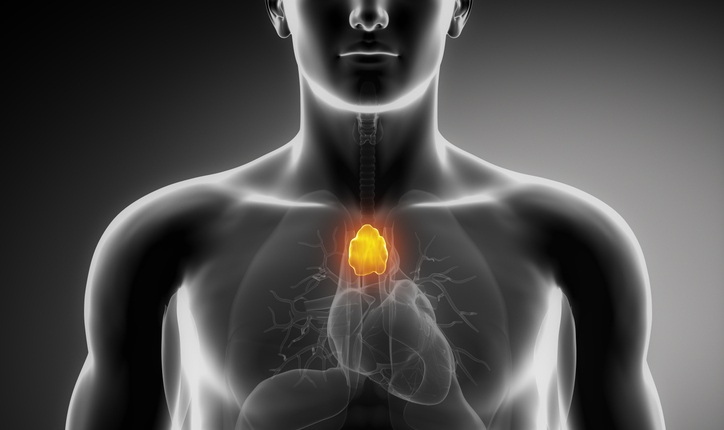
Study Links Thymus Radiation Dose to Poorer Outcomes in Lung Cancer
Collateral radiation to the thymus during therapy for non-small cell lung cancer (NSCLC) may compromise antitumor immunity and disease control. The thymus is a small immune organ in the upper chest that... Read more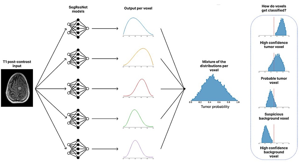
Health IT
view channel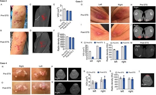
Imaging Tool Quantifies Calcinosis Cutis Volume to Monitor Treatment
Calcinosis cutis involves calcium deposits in the skin or soft tissues that can develop anywhere in the body, causing pain, impaired mobility, and disability. Lesions along the spine or buttocks can make... Read more
Digital Symptom Monitoring Supports More Personalized Breast Cancer Care
Metastatic breast cancer, in which tumors spread to other organs, is incurable but increasingly managed with modern systemic therapies. As more treatments shift from hospital infusions to oral or at-home... Read more

Point of Care
view channel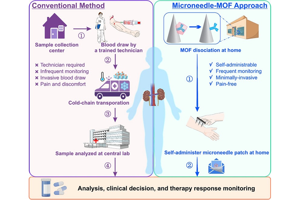
Microneedle Patch Enables At-Home Monitoring of Acute Kidney Injury
Kidney disease is often silent in its early stages, making timely detection difficult outside clinical settings. Early biomarkers such as neutrophil gelatinase–associated lipocalin can require blood draws... Read more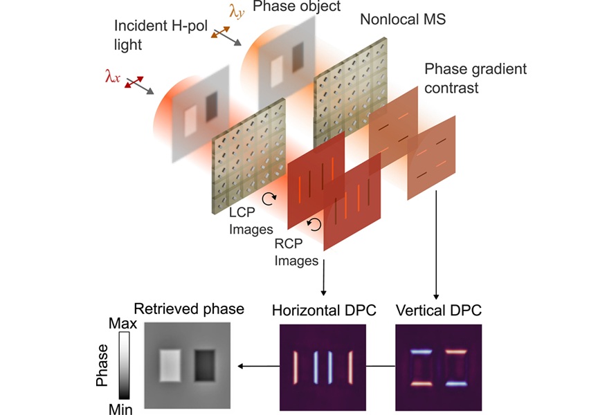
Ultrathin Metasurface Could Bring Quantitative Phase Imaging to Portable Devices
Point-of-care imaging often depends on bulky optical systems that have difficulty visualizing nearly transparent cells and tissues without staining. These limitations can slow diagnosis and restrict use... Read more
Business
view channel
First U.S. Commercial Cases Demonstrate New Approach to Transseptal Access
Transseptal access to the left atrium is critical in catheter-based cardiac procedures, where direct visualization and precise positioning are essential. Clinicians also aim to reduce fluoroscopy use and... Read more