



Google Shows AI Can Predict Lung Cancer from CT Scans
|
By HospiMedica International staff writers Posted on 01 Jun 2019 |


|
Google LLC (Mountain View, CA, USA) has shared new research demonstrating how artificial intelligence (AI) can predict lung cancer to boost the chances of survival for people at risk across the world.
Since 2017, Google has been exploring how AI can be used to address the challenges in screening people at a high-risk for lung cancer with lower dose CT screening that leads to unclear diagnosis, subsequent unnecessary procedures, and financial costs. Google used advances in 3D volumetric modeling along with datasets from its partners for modeling lung cancer prediction and laying the groundwork for future clinical testing.
Generally, radiologists go through hundreds of 2D images within a single CT scan with cancer being miniscule and hard to spot. Google researchers created a model that can generate the overall lung cancer malignancy prediction (viewed in 3D volume) as well as identify subtle malignant tissue in the lungs (lung nodules). The model can also factor in information from previous scans, which can be useful in predicting lung cancer risk as the growth rate of suspicious lung nodules can be an indicator of malignancy.
The researchers leveraged 45,856 de-identified chest CT screening cases and validated the results with a second dataset and also compared their results against six US board-certified radiologists. They found that when using a single CT scan for diagnosis, their model performed on par or better than the six radiologists and detected 5% more cancer cases while reducing false-positive exams by more than 11% as compared to unassisted radiologists in the study. Google’s approach achieved an AUC (a common metric used in machine learning that provides an aggregate measure for classification performance) of 94.4%.
With only 2-4% of eligible patients in the US being currently screened for lung cancer, Google’s research demonstrates the potential for AI to increase accuracy as well as consistency, which could help accelerate the adoption of lung cancer screening globally. Google now plans to conduct further studies to assess its impact and utility in clinical practice. It is collaborating with Google Cloud Healthcare and Life Sciences team to serve the model through the Cloud Healthcare API and is holding discussions with its partners across the world to continue additional clinical validation research and deployment.
Related Links:
Google




Since 2017, Google has been exploring how AI can be used to address the challenges in screening people at a high-risk for lung cancer with lower dose CT screening that leads to unclear diagnosis, subsequent unnecessary procedures, and financial costs. Google used advances in 3D volumetric modeling along with datasets from its partners for modeling lung cancer prediction and laying the groundwork for future clinical testing.
Generally, radiologists go through hundreds of 2D images within a single CT scan with cancer being miniscule and hard to spot. Google researchers created a model that can generate the overall lung cancer malignancy prediction (viewed in 3D volume) as well as identify subtle malignant tissue in the lungs (lung nodules). The model can also factor in information from previous scans, which can be useful in predicting lung cancer risk as the growth rate of suspicious lung nodules can be an indicator of malignancy.
The researchers leveraged 45,856 de-identified chest CT screening cases and validated the results with a second dataset and also compared their results against six US board-certified radiologists. They found that when using a single CT scan for diagnosis, their model performed on par or better than the six radiologists and detected 5% more cancer cases while reducing false-positive exams by more than 11% as compared to unassisted radiologists in the study. Google’s approach achieved an AUC (a common metric used in machine learning that provides an aggregate measure for classification performance) of 94.4%.
With only 2-4% of eligible patients in the US being currently screened for lung cancer, Google’s research demonstrates the potential for AI to increase accuracy as well as consistency, which could help accelerate the adoption of lung cancer screening globally. Google now plans to conduct further studies to assess its impact and utility in clinical practice. It is collaborating with Google Cloud Healthcare and Life Sciences team to serve the model through the Cloud Healthcare API and is holding discussions with its partners across the world to continue additional clinical validation research and deployment.
Related Links:

Gold Member
12-Channel ECG
CM1200B
Medical-Grade Display
HL2316SHTB
Breast Imaging Monitor
Barco Coronis Onelook MDMC-32133 32MP
Latest AI News
- AI Tool Improves Smartphone Detection of Abnormal Pediatric Lung Sounds
- AI Tool Detects Coronary Heart Disease from Face and Tongue Images
- Optical Imaging System Helps Predict Chemotherapy Response in Cancer
- AI Tool Predicts Long-Term Cardiovascular Risk from Sleep Study ECG
- Predictive Model Supports Growth Hormone Treatment Planning in Idiopathic Short Stature
- AI Model Predicts Prolonged Sitting in Chronic Pelvic Pain
- Autonomous AI Helps Streamline Urgent Skin Cancer Referrals
- Virtual Ultrasound Images Support Development of Cardiovascular Imaging Tools
- Explainable AI Tool Improves Brain Disorder Screening from 4D fMRI
- New Benchmark Highlights Evidence Gaps in AI Responses to Clinical Questions
- AI-Powered ECG Model Gains FDA Authorization for Emergency Cardiac Assessment
- AI Improves Non-Contrast CT Interpretation for Time-Sensitive Stroke Assessment
- Explainable AI Tool Identifies High-Risk Patients Before Heart Attack Reperfusion
- AI Tools Show Potential to Detect Subtle Signs of Interval Breast Cancer
Channels
Artificial Intelligence
view channel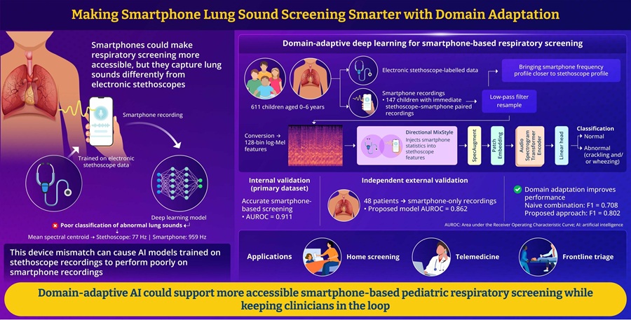
AI Tool Improves Smartphone Detection of Abnormal Pediatric Lung Sounds
Respiratory disease in children remains a major clinical concern worldwide. Abnormal lung sounds can indicate pediatric respiratory illness, but standard auscultation usually requires an in-person examination... Read more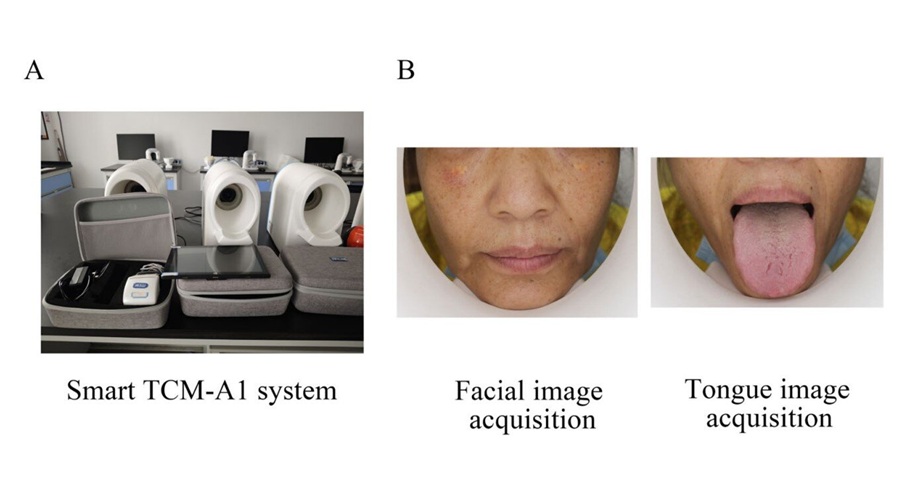
AI Tool Detects Coronary Heart Disease from Face and Tongue Images
Coronary heart disease (CHD) develops when fatty plaques narrow the arteries supplying the heart muscle, restricting blood flow and sometimes first presenting as chest pain, myocardial infarction, or another... Read more

Critical Care
view channel
Smart Pulse Oximetry Supports Earlier Recognition of Arrhythmias and Respiratory Risk
On general care floors, respiratory compromise and arrhythmias can develop between routine checks. Atrial fibrillation often goes undiagnosed in older adults, while opioid-induced respiratory depression... Read more
Proposed Risk Score Prioritizes Sickest Patients for Heart Transplant
Donor hearts remain extremely scarce in the U.S. transplant system, with one in five candidates dying or being removed from the waiting list before receiving an organ. Current allocation rules may also... Read more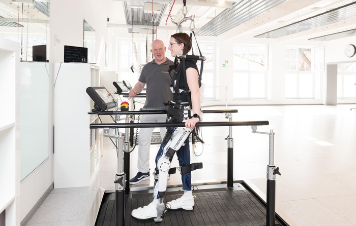

Surgical Techniques
view channel
3D Printing Method Could Enable More Flexible Cardiovascular Implants
Minimally invasive transcatheter devices must compress into small catheters and recover their intended shape after deployment, creating a design challenge for cardiovascular implants with complex, patient-specific... Read more
FDA Approves Bioabsorbable Patch Technology for Large-Bore Vascular Closure
Structural heart and endovascular procedures often require large-bore arterial access, increasing the need for reliable vascular closure after intervention. Closure systems must achieve hemostasis while... Read more
Robotic Approach Enables Minimally Invasive Repair of Hard-to-Reach Abdominal Hernias
Lateral abdominal wall hernias are uncommon defects that develop along the side of the abdomen and can be difficult to repair because of their proximity to the ribs, pelvis, muscles, nerves, and blood vessels.... Read more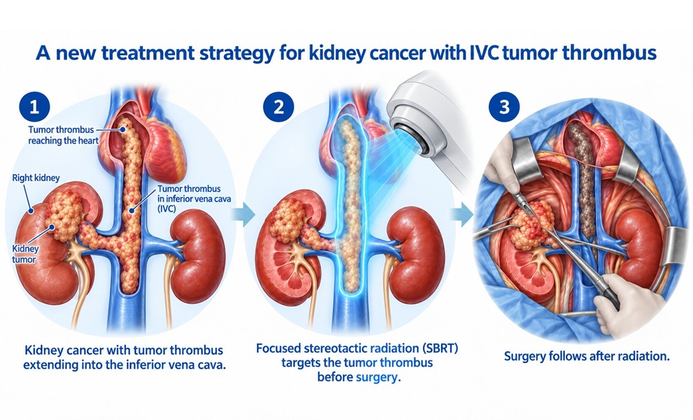
Patient Care
view channel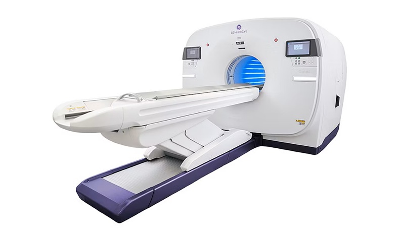
Total-Body PET/CT Scanner Enables One-Minute Diagnostic-Quality Cancer Imaging
Positron emission tomography/computed tomography (PET/CT) combines radiotracer-based functional imaging with anatomical detail to diagnose, stage, and monitor cancer. Conventional scanners image the body... Read more
AI Platform Personalizes Support Between Visits for Cancer Survivors
Cancer survivors often manage persistent side effects after treatment, including fatigue, anxiety, uncertainty, symptoms, stress, and questions between appointments. Clinical teams need ways to maintain... Read more
At-Home Neuromodulation Shows Promise in Selected Patients With Hypertension
High blood pressure is a major risk factor for cardiovascular disease, including heart attack and stroke. Many patients receiving treatment for hypertension still remain above recommended blood pressure... Read more
EHR Model Flags High-Risk Periods in Patients with Metastatic Breast Cancer
Determining when patients with metastatic breast cancer are nearing the end of life remains difficult, often leading to intensive interventions with limited benefit. Prognostic uncertainty can delay goals-of-care... Read moreHealth IT
view channel
EHR Alerts Support Dosing, Sedation, and Shock Recognition in Cardiac Care
Electronic health record alerts can add to clinician workload when they are poorly targeted. In critical cardiac care, however, delayed decisions can affect treatment timing, sedation exposure, and escalation... Read more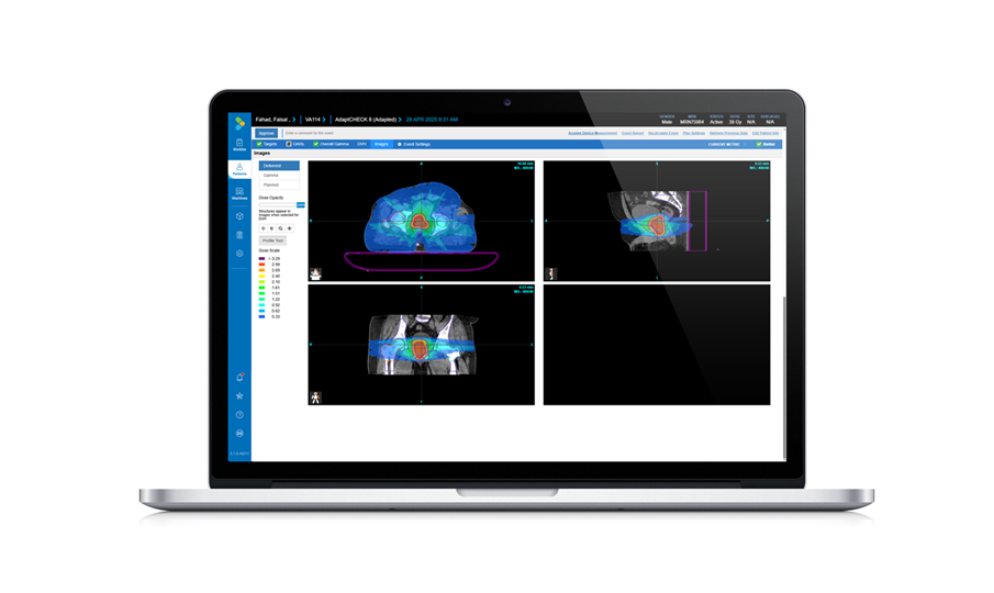
Adaptive Radiation QA Software Gains Validation for NeoArc Treatments
Adaptive radiation therapy allows clinicians to adjust treatment plans to daily anatomical changes such as tumor shrinkage, weight loss, or organ movement, improving precision while limiting exposure to... Read more

Point of Care
view channel
Continuous Glucose Monitoring Identifies Cardiometabolic Risk in Adults Without Diabetes
Dysglycemia—abnormal blood glucose regulation—can fluctuate throughout the day and often escape conventional screening. Clinicians typically rely on fasting plasma glucose and hemoglobin A1c, which offer... Read more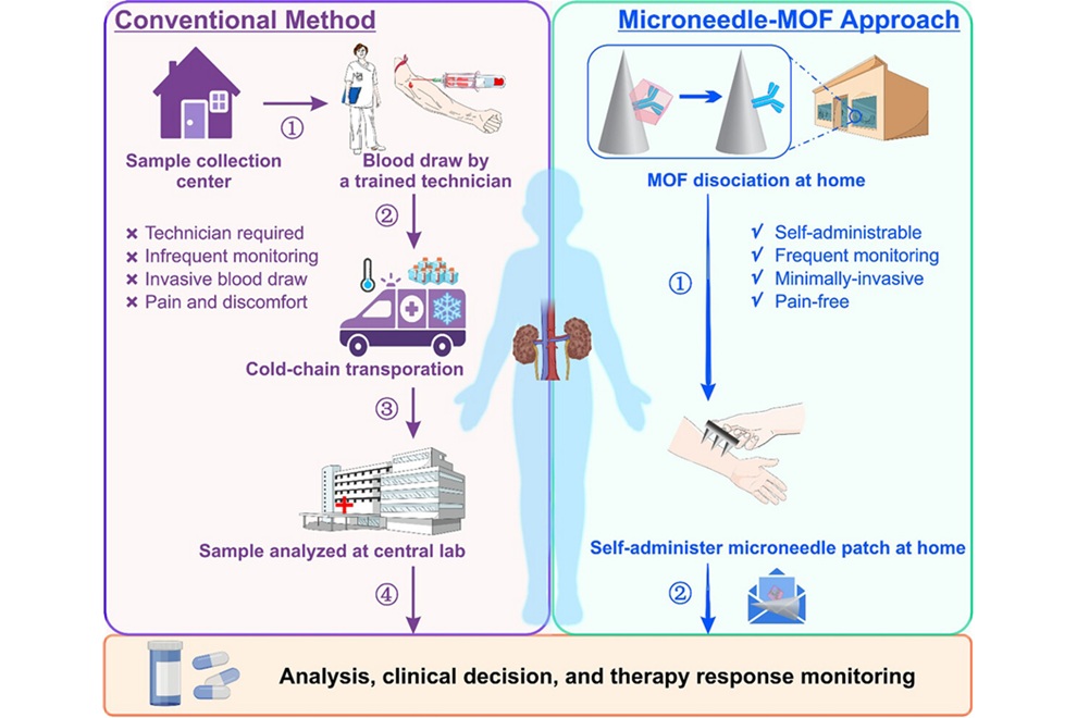
Microneedle Patch Enables At-Home Monitoring of Acute Kidney Injury
Kidney disease is often silent in its early stages, making timely detection difficult outside clinical settings. Early biomarkers such as neutrophil gelatinase–associated lipocalin can require blood draws... Read more
Business
view channel
Steris to Expand Surgical Safety Portfolio With $2.1 Billion Xodus Medical Deal
Steris has signed a definitive agreement to acquire Xodus Medical, a specialty surgical products business based in Pennsylvania, for $2.1 billion at closing. The acquisition will expand Steris’s healthcare... Read more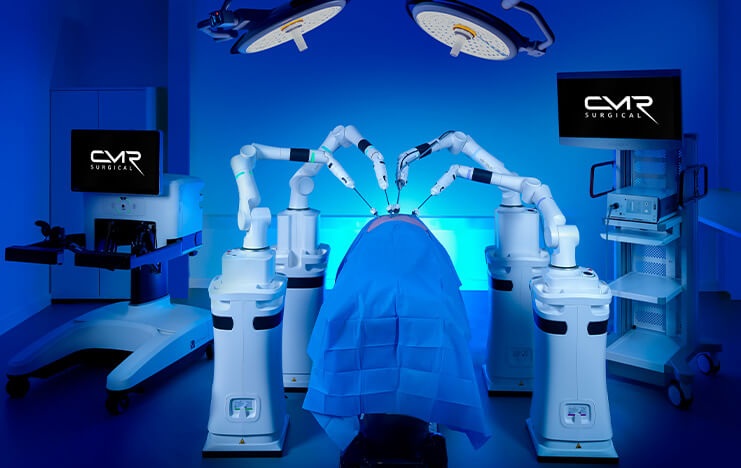
Stryker Enters Soft-Tissue Robotics Through Strategic Alliance With CMR Surgical
Stryker (Portage, MI, USA) and CMR Surgical (Cambridge, UK) have announced a strategic alliance centered on the Versius Surgical Robotic portfolio. The agreement includes an approximately USD 400 million... Read more
Terumo Neuro Acquisition Advances Liquid Embolic Technology for Chronic Subdural Hematoma
Terumo Neuro (Aliso Viejo, CA, USA) signed an agreement to acquire Arsenal Medical Inc. (Waltham, MA, USA), a privately held, clinical-stage biomaterials company. The proposed acquisition would add NeoCast,... Read more