


Approximation Kit Closes Microdiscectomy Disc Defects
|
By HospiMedica International staff writers Posted on 29 Jul 2019 |


|
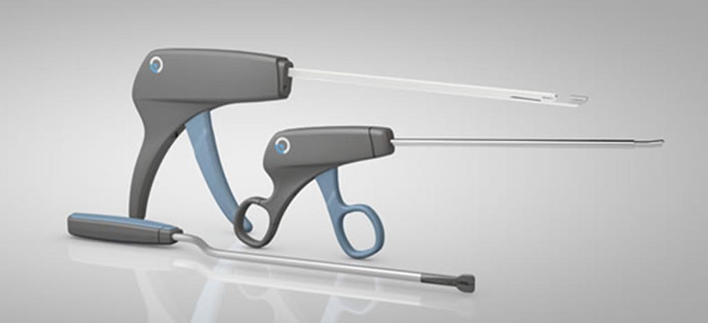
Image: The AnchorKnot Tissue Approximation Kit (Photo courtesy of Anchor Orthopedics).
A novel tissue sparing approximation system minimizes residual defects during herniated disc repair and improves procedural outcomes.
The Anchor Orthopedics XT (Mississauga, Canada) AnchorKnot Tissue Approximation Kit is a set of surgical spinal devices that enable orthopedic surgeons and neurosurgeons to close tissue defects during minimally invasive microdiscectomy of herniated intervertebral discs in the lumbar spine, allowing surgical decompression with minimization of the annular incision and limiting the removal of tissue to include only the free fragments impinging on the nerve root. The kit is comprised of three sterile, single use devices: the Suture Passer, the Knot Pusher, and the Suture Cutter.
The Suture Passer is designed for passing a preloaded non-absorbable ultra-high molecular weight (UHMW) polyethylene surgical suture through soft tissue. The Knot Pusher is a hand-held grip device, with a stainless steel shaft and a slotted tip that is used to push the pre-tied knot down to the tissue level. The Suture Cutter is used to cut the suture once the knot is secured in place. The deployment mechanism of the AnchorKnot tissue approximation kit is designed for simplicity, despite the difficult to reach anatomical location; on average, about five minutes of usage are needed during the procedure.
“Microdiscectomy surgery allows a patient to return to a more healthy state, faster and with less pain; the AnchorKnot kit allows us to repair tissue defects with an elegant device,” said Nilesh Patel, MD, of Michigan Spine Clinic (Brownstown, USA). “This technology represents the next evolution in minimally invasive herniated disc repair. We are always looking to reduce the size of the tissue defect and we see it as a critical step towards our goal of and improving procedure outcomes.”
A lumbar disc herniation is a tear in the outer, fibrous ring (annulus fibrosus) of an intervertebral disc that allows part of, or the entire, soft central portion to be forced out from between two vertebrae, causing back pain, and can also push on nerve roots causing pain, numbness and tingling, as well as weakness in one or both legs. Symptomatic lumbar disc herniation affects 1-2% of the population at some point in their lives, usually between 30-40 years of age, and is one of the most common and debilitating conditions of the spine.
Related Links:
Anchor Orthopedics XT



The Anchor Orthopedics XT (Mississauga, Canada) AnchorKnot Tissue Approximation Kit is a set of surgical spinal devices that enable orthopedic surgeons and neurosurgeons to close tissue defects during minimally invasive microdiscectomy of herniated intervertebral discs in the lumbar spine, allowing surgical decompression with minimization of the annular incision and limiting the removal of tissue to include only the free fragments impinging on the nerve root. The kit is comprised of three sterile, single use devices: the Suture Passer, the Knot Pusher, and the Suture Cutter.
The Suture Passer is designed for passing a preloaded non-absorbable ultra-high molecular weight (UHMW) polyethylene surgical suture through soft tissue. The Knot Pusher is a hand-held grip device, with a stainless steel shaft and a slotted tip that is used to push the pre-tied knot down to the tissue level. The Suture Cutter is used to cut the suture once the knot is secured in place. The deployment mechanism of the AnchorKnot tissue approximation kit is designed for simplicity, despite the difficult to reach anatomical location; on average, about five minutes of usage are needed during the procedure.
“Microdiscectomy surgery allows a patient to return to a more healthy state, faster and with less pain; the AnchorKnot kit allows us to repair tissue defects with an elegant device,” said Nilesh Patel, MD, of Michigan Spine Clinic (Brownstown, USA). “This technology represents the next evolution in minimally invasive herniated disc repair. We are always looking to reduce the size of the tissue defect and we see it as a critical step towards our goal of and improving procedure outcomes.”
A lumbar disc herniation is a tear in the outer, fibrous ring (annulus fibrosus) of an intervertebral disc that allows part of, or the entire, soft central portion to be forced out from between two vertebrae, causing back pain, and can also push on nerve roots causing pain, numbness and tingling, as well as weakness in one or both legs. Symptomatic lumbar disc herniation affects 1-2% of the population at some point in their lives, usually between 30-40 years of age, and is one of the most common and debilitating conditions of the spine.
Related Links:
Anchor Orthopedics XT

Gold Member
Blood Gas Analyzer
i-Check200
Digital Radiography System (Ceiling Free)
Digix CF Series
Desk Aneroid Sphyg
Diagnostix 750D+
Latest Surgical Techniques News
- FDA Approves First Sirolimus Drug-Eluting Balloon for Coronary In-Stent Restenosis
- Light-Activated Nanoparticles May Offer New Approach to Retinal Prostheses
- Handheld Infrared Imaging Device Supports Tumor Margin Assessment During Surgery
- Needle-Free Intrauterine Anesthesia Technique Reduces Pain During IUD Insertion
- Robotic Platform Advances Fetoscopic Treatment of Twin-to-Twin Transfusion Syndrome
- Patient-Specific AI Tool Aligns X-Rays With 3D Scans For Surgical Navigation
- FDA Clears Integrated Platform for Real-Time Cardiac Imaging and Catheter Guidance
- Hydrogel Dermal Supports Healing of Complex Wounds After Cancer Surgery
- New Microcatheter Enhances Lesion Crossing in Complex Coronary Interventions
- Integrated PET/MRI and EEG Improves Presurgical Lesion Localization in Pediatric Focal Epilepsy
- Integrated Perioperative Care Model Improves Recovery in Older Surgical Patients
- New Transvaginal Transducer Improves Imaging for Gynecology and Early Obstetrics
- FDA Approves Abbott Dual-Energy Catheter for Complex AFib Cases
- Mobile 3-in-1 Imaging System Advances Intraoperative Navigation
- First Thyroid Cancer-Specific Anxiety Scale Supports Patient-Centered Care
- Tissue-Mimicking Scaffold Could Improve Rotator Cuff Repair
Channels
Artificial Intelligence
view channel
AI Improves Non-Contrast CT Interpretation for Time-Sensitive Stroke Assessment
Acute ischemic stroke occurs when a blood vessel in the brain becomes blocked, requiring rapid diagnosis to enable timely reperfusion therapy. Emergency departments often use computed tomography angiography... Read more
AI-Powered ECG Model Gains FDA Authorization for Emergency Cardiac Assessment
Powerful Medical’s PMcardio Queen of Hearts (ACS AI ECG Model) has received U.S. Food and Drug Administration (FDA) De Novo authorization, announced on September 15, 2026. The model is designed to identify... Read more
Critical Care
view channel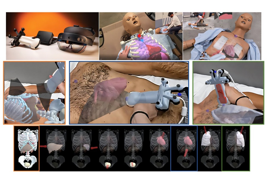
Anatomical Guidance System Improves Trauma Ultrasound Acquisition by Novice Users
Focused assessment with sonography for trauma (FAST) helps clinicians detect free fluid and signs of internal bleeding in injured patients, but obtaining reliable images can be difficult for inexperienced operators.... Read more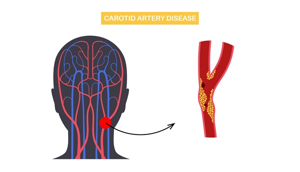
AI-Enhanced Handheld Ultrasound Improves Carotid Plaque Detection
Handheld ultrasound can miss small or faint carotid plaque, creating uncertainty in community screening. This gap makes it harder for frontline clinicians to decide who needs confirmatory imaging, closer... Read more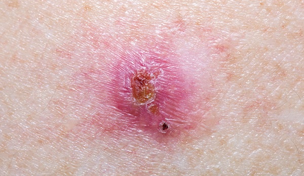
Noninvasive Imaging Approach Aims to Detect Basal Cell Carcinoma Before It Becomes Visible
Basal cell carcinoma is the most common form of skin cancer and can damage nearby structures such as the nose or eyes when it develops on the face. Diagnosis typically depends on visible skin changes,... Read more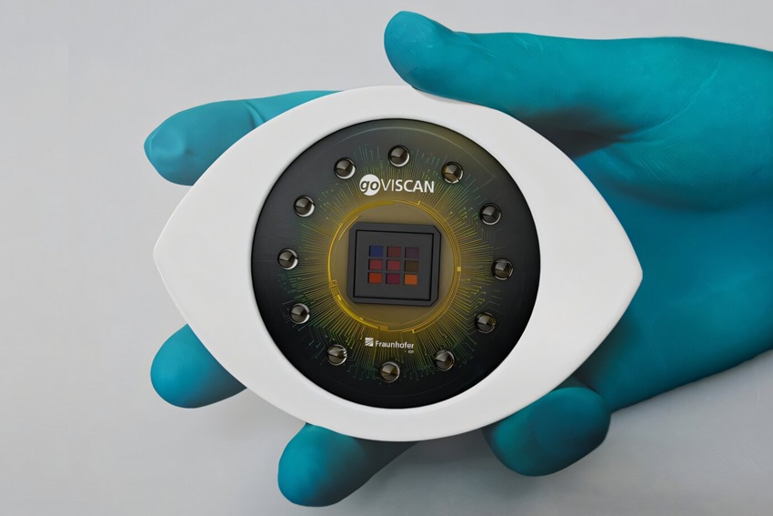
Patient Care
view channel
EHR Model Flags High-Risk Periods in Patients with Metastatic Breast Cancer
Determining when patients with metastatic breast cancer are nearing the end of life remains difficult, often leading to intensive interventions with limited benefit. Prognostic uncertainty can delay goals-of-care... Read more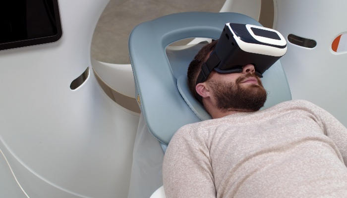
Virtual Reality Helps Claustrophobic Patients Complete Cardiac PET/CT Scans
Anxiety and claustrophobia—intense fear of confined spaces—often prevent patients from completing cardiac positron emission tomography/computed tomography (PET/CT). Aborted scans delay diagnosis, consume... Read more

Medical Imaging
view channel
New MRI Approach Captures Rapidly Decaying Tissue Signals
MRI measures signals generated as excited proton spins return toward equilibrium. However, some tissue signals decay so rapidly that conventional systems can miss them during the brief interval after excitation,... Read more
AI Tool Integrates MRI and Ultrasound to Detect Endometriosis
Endometriosis is a chronic condition in which tissue similar to the uterine lining grows outside the uterus, causing pain, heavy bleeding, and infertility. Diagnosis often depends on surgical visualization,... Read more
Health IT
view channel
New Tablet Platform Extends Stroke Neurorehabilitation Beyond the Clinic
Continuity and intensity of neurorehabilitation after stroke are critical for functional recovery, but access to ongoing therapy at home often remains limited. Hospitals and outpatient programs therefore... Read more
Smartphone App Improves Blood Pressure Control in Hypertension
Hypertension, or high blood pressure, is a major modifiable driver of cardiovascular, renal, and cerebrovascular morbidity. Despite effective therapies, many adults fail to achieve control, leading to... Read more

Point of Care
view channel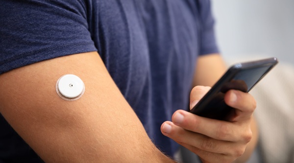
Continuous Glucose Monitoring Identifies Cardiometabolic Risk in Adults Without Diabetes
Dysglycemia—abnormal blood glucose regulation—can fluctuate throughout the day and often escape conventional screening. Clinicians typically rely on fasting plasma glucose and hemoglobin A1c, which offer... Read more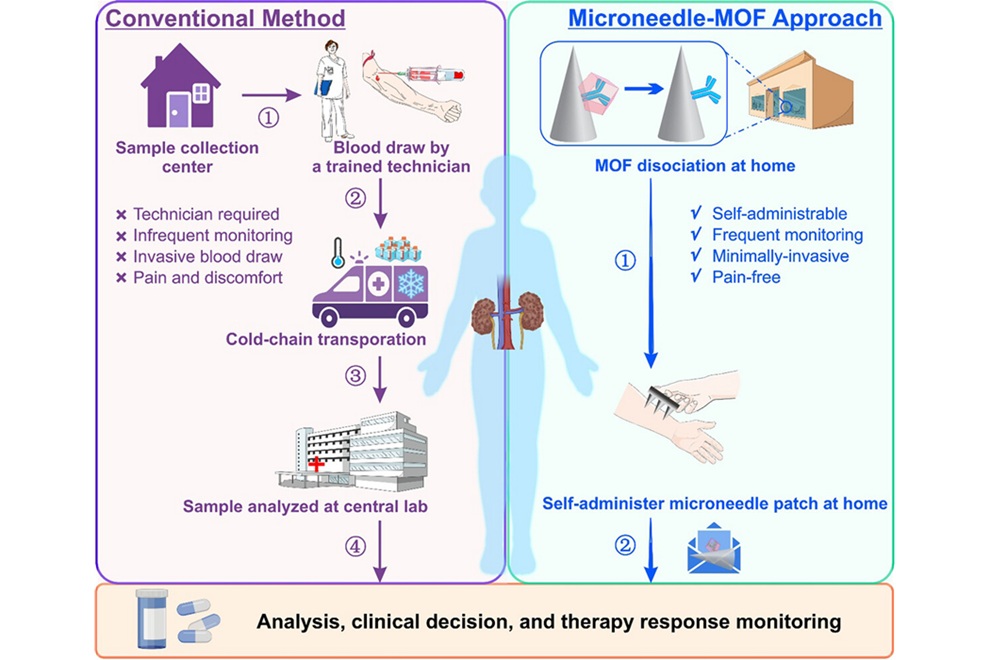
Microneedle Patch Enables At-Home Monitoring of Acute Kidney Injury
Kidney disease is often silent in its early stages, making timely detection difficult outside clinical settings. Early biomarkers such as neutrophil gelatinase–associated lipocalin can require blood draws... Read more

Business
view channel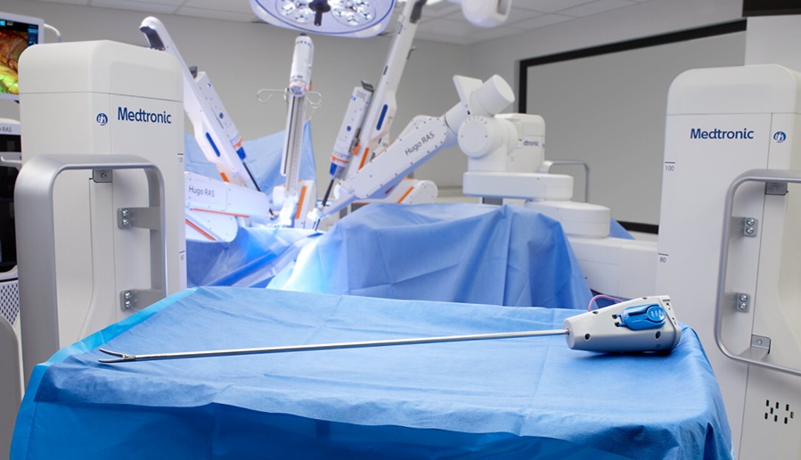
Medtronic Receives FDA Clearance for Vessel-Sealing Instrument for Robotic Surgery
As robotic-assisted surgery expands across U.S. hospitals, teams increasingly seek energy instruments with the familiarity and performance of tools used in open and laparoscopic procedures.... Read more
Robotic Single-Port System Gains CE Mark for Transvaginal Gynecologic Procedures
Intuitive (Sunnyvale, CA, USA) announced that it has received CE mark approval for use of the da Vinci SP Single Port surgical system in transvaginal gynecologic procedures, marking the first such indication... Read more