


Robotic System Simplifies the Spinal Surgery Process
|
By HospiMedica International staff writers Posted on 02 Jun 2021 |


|
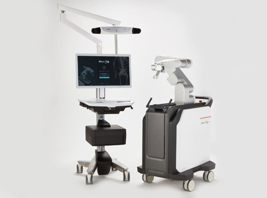
Image: The CUVIS-Spine spinal surgery robot main console (L) and robotic arm (R) (Photo courtesy of Curexo)
A highly accurate one-step surgical tool with a real-time tracking sensor provides practitioners with fast, simple, and precise pedicle screw insertion.
The Curexo (Seoul, South Korea) CUVIS-Spine is a next-gen spinal surgery robot that guides the insertion of a pedicle screw according to the surgery plan. The system includes uses a high precision robot arm and wireless, one-step navigation based on a real-time GPS-like sensor that provide precise, safe, and faster surgery, when compared to traditional manual placement. It also minimizes X-ray radiation exposure of both patients and medical staff, as only two slices (AP and LL) are needed for plan the pedicle screws entry point, target point, and route.
Both two-dimensional X-ray filming (via a C-arm) and three -dimensional filming (with an O-arm) are available, expanding clinician’s options. The wireless one-step navigation and robot arms allow multiple steps, such as needling, K-wire manipulation, dilation, and tapping) to be performed using just one tool, reducing surgery duration greatly. In order to prevents surgical instruments from skiving, an optical tracking system (OTS) provides accurate data on the lateral force affecting the arm and pedicle screw in real-time.
“The spinal surgery robot 'CUVIS-spine' that we have developed and manufactured with our technology has acquired FDA (US)'s licensing. This is the 3rd license that we have acquired, followed by Korea and Europe, and now we can sell our solution in all countries including the US which is the biggest medical market." He also said that "CUREXO plans to focus on our medical robots sales, including 'CUVIS-joint', 'CUVIS-spine' and 'Morning Walk' to not only the Korean market but also to advanced medical markets such as the US and Europe."
“CUVIS-Spine had successfully carried out the insertion of pedicle screws on a patient with spinal stenosis and degenerative spondylolisthesis,” said Jae-Joon Lee, CEO of Curexo, following the first successful spinal robotic surgery at Severance Hospital (Sinchon, South Korea). “After the surgery, image examinations had proven the fixation of the screws as originally planned, and the patient was able to leave the hospital on the fifth day with improvements.”
“I expect that Korean robots will be able to enter the market by accumulating track records and creating clinical evidence in the field of spinal surgical robots, which is expanding globally," said Seong Yi, MD, of the department of neurosurgery at Severance Hospital, who participated in the development of CUVIS-Spine. “Clinical data of spinal surgical robots, including safety, validity, and radiation safety level will contribute to the development and globalization of the domestic surgical robot industry.”
Pedicle screws provide a means of gripping a spinal segment. The screws themselves do not fixate the spinal segment, but act as firm anchor points that can then be connected with a rod. The screws are placed at two or three consecutive spine segments and then a short rod is used to connect the screws; this construct prevents motion at the segments that are being fused. After the bone graft grows, the screws and rods are no longer needed for stability and may be safely removed. However, most surgeons do not recommend removal unless the pedicle screws cause discomfort for the patient.
Related Links:
Curexo




The Curexo (Seoul, South Korea) CUVIS-Spine is a next-gen spinal surgery robot that guides the insertion of a pedicle screw according to the surgery plan. The system includes uses a high precision robot arm and wireless, one-step navigation based on a real-time GPS-like sensor that provide precise, safe, and faster surgery, when compared to traditional manual placement. It also minimizes X-ray radiation exposure of both patients and medical staff, as only two slices (AP and LL) are needed for plan the pedicle screws entry point, target point, and route.
Both two-dimensional X-ray filming (via a C-arm) and three -dimensional filming (with an O-arm) are available, expanding clinician’s options. The wireless one-step navigation and robot arms allow multiple steps, such as needling, K-wire manipulation, dilation, and tapping) to be performed using just one tool, reducing surgery duration greatly. In order to prevents surgical instruments from skiving, an optical tracking system (OTS) provides accurate data on the lateral force affecting the arm and pedicle screw in real-time.
“The spinal surgery robot 'CUVIS-spine' that we have developed and manufactured with our technology has acquired FDA (US)'s licensing. This is the 3rd license that we have acquired, followed by Korea and Europe, and now we can sell our solution in all countries including the US which is the biggest medical market." He also said that "CUREXO plans to focus on our medical robots sales, including 'CUVIS-joint', 'CUVIS-spine' and 'Morning Walk' to not only the Korean market but also to advanced medical markets such as the US and Europe."
“CUVIS-Spine had successfully carried out the insertion of pedicle screws on a patient with spinal stenosis and degenerative spondylolisthesis,” said Jae-Joon Lee, CEO of Curexo, following the first successful spinal robotic surgery at Severance Hospital (Sinchon, South Korea). “After the surgery, image examinations had proven the fixation of the screws as originally planned, and the patient was able to leave the hospital on the fifth day with improvements.”
“I expect that Korean robots will be able to enter the market by accumulating track records and creating clinical evidence in the field of spinal surgical robots, which is expanding globally," said Seong Yi, MD, of the department of neurosurgery at Severance Hospital, who participated in the development of CUVIS-Spine. “Clinical data of spinal surgical robots, including safety, validity, and radiation safety level will contribute to the development and globalization of the domestic surgical robot industry.”
Pedicle screws provide a means of gripping a spinal segment. The screws themselves do not fixate the spinal segment, but act as firm anchor points that can then be connected with a rod. The screws are placed at two or three consecutive spine segments and then a short rod is used to connect the screws; this construct prevents motion at the segments that are being fused. After the bone graft grows, the screws and rods are no longer needed for stability and may be safely removed. However, most surgeons do not recommend removal unless the pedicle screws cause discomfort for the patient.
Related Links:
Curexo

Gold Member
Neonatal Heel Incision Device
Tenderfoot
Surgical System
Stealth AXiS
Fetal Monitor
BT-380
Latest Surgical Techniques News
- New Microcatheter Enhances Lesion Crossing in Complex Coronary Interventions
- Integrated PET/MRI and EEG Improves Presurgical Lesion Localization in Pediatric Focal Epilepsy
- Integrated Perioperative Care Model Improves Recovery in Older Surgical Patients
- New Transvaginal Transducer Improves Imaging for Gynecology and Early Obstetrics
- FDA Approves Abbott Dual-Energy Catheter for Complex AFib Cases
- Mobile 3-in-1 Imaging System Advances Intraoperative Navigation
- First Thyroid Cancer-Specific Anxiety Scale Supports Patient-Centered Care
- Tissue-Mimicking Scaffold Could Improve Rotator Cuff Repair
- Augmented Reality System Integrates Intraoperative Data for Hip Arthroscopy
- Nanobone Material Activates Natural Repair Signals to Regrow Bone
- First Robotic Dual Liver-Kidney Transplant Expands Minimally Invasive Transplant Surgery
- Functional Coronary Angiography Improves Outcomes in STEMI With Multivessel Disease
- Researchers Advance Novel Therapy for Hard-to-Heal Diabetic Foot Wounds
- Robotic System Aims to Expand Access to Stroke Thrombectomy
- Biodegradable Gastric Stent Could Eliminate Repeat Procedures After Bariatric Surgery
- Robot-Assisted Surgery Shows Advantages Over Laparoscopy in Gastric Cancer
Channels
Artificial Intelligence
view channel
Explainable AI Tool Identifies High-Risk Patients Before Heart Attack Reperfusion
Intramyocardial hemorrhage is bleeding within the heart muscle that can occur after blood flow is restored during heart attack treatment. It affects about 40% of patients treated for ST-segment elevation... Read more
AI Tools Show Potential to Detect Subtle Signs of Interval Breast Cancer
Interval breast cancers, diagnosed after a negative screening mammogram and before the next scheduled exam, remain an important measure of screening performance and are often more aggressive.... Read more

Critical Care
view channel
Wearable Microneedle Device Aims to Enable Remotely Managed Pain Relief
Chronic pain often forces patients to rely on repeat clinic visits or prolonged analgesic use, which carries risks of side effects and dependency. Opioids for severe pain can lead to tolerance and misuse,... Read more
Octopus-Inspired Patch Improves Adhesion for Transdermal Drug Delivery
Transdermal drug delivery requires secure contact between a patch and dynamically moving skin. Everyday bending and stretching can open microgaps that degrade adhesion and reduce dose consistency, limiting... Read more
Patient Care
view channel
Virtual Reality Helps Claustrophobic Patients Complete Cardiac PET/CT Scans
Anxiety and claustrophobia—intense fear of confined spaces—often prevent patients from completing cardiac positron emission tomography/computed tomography (PET/CT). Aborted scans delay diagnosis, consume... Read more
Weighted Blankets Reduce Anxiety in Hospitalized Adults
Anxiety is common among hospitalized adults and can impair recovery, disrupt sleep, and worsen mental health. Although clinicians often use nonpharmacologic strategies, bedside options that act quickly... Read more

Medical Imaging
view channel
Noninvasive Optical Test Detects Hidden Tissue Damage in Pelvic Organ Prolapse
Pelvic organ prolapse (POP) occurs when weakened pelvic-support tissues allow the bladder, uterus, or rectum to descend. Although it affects millions of women, the condition remains under-characterized... Read more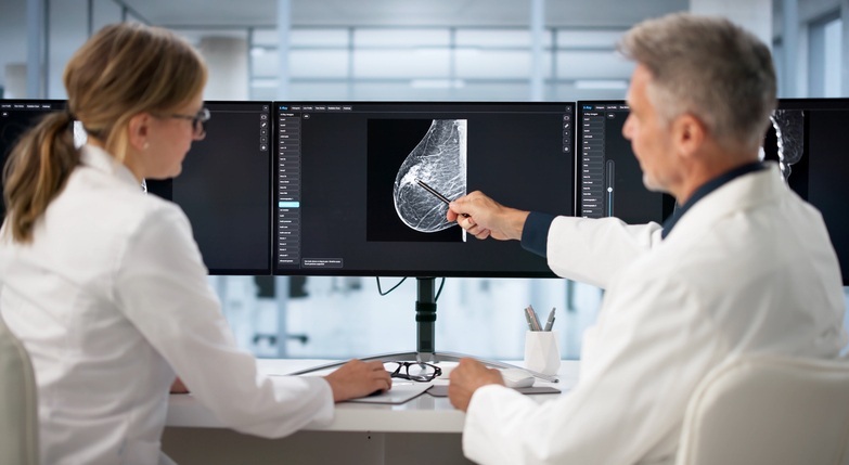
AI Imaging Model Predicts Five-Year Breast Cancer Risk from 3D Mammograms
Breast cancer screening programs often struggle to stratify women by near-term risk, which can lead to missed cancers or unnecessary testing. Forecasting who will develop disease within five years is difficult... Read more
Spleen Imaging Signatures May Reveal Hidden Coronary Artery Disease Risk
Coronary artery disease (CAD) remains a leading cause of death despite major gains in managing traditional risk factors. Clinicians still struggle to quantify residual risk and the upstream biology that... Read more
FDA Clears Automated Musculoskeletal Ultrasound Platform for Arthritis Assessment
Musculoskeletal ultrasound is central to evaluating inflammatory arthritis and other joint conditions, but access is limited by workforce shortages and variability in image acquisition. In the United States,... Read moreHealth IT
view channel
Registry-Driven Digital Outreach Boosts Use of Heart Failure Medicines
Heart failure affects more than 64 million people worldwide and is linked to high mortality, reduced quality of life, and heavy healthcare use. Although sodium-glucose cotransporter 2 inhibitors are proven... Read more
Nurse-Coordinated Care Model Improves Heart Failure Outcomes
Heart failure imposes heavy mortality, morbidity, and cost burdens worldwide, yet proven therapies remain underused in routine practice. This inadequate uptake contributes to preventable hospitalizations... Read more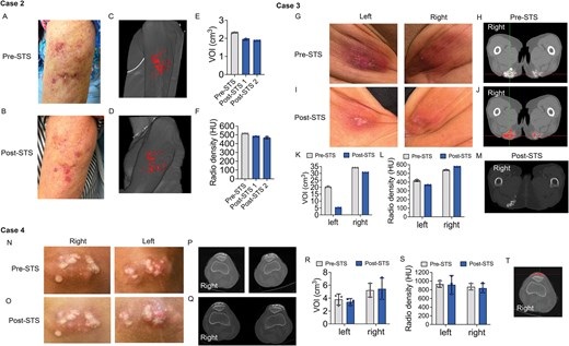

Point of Care
view channel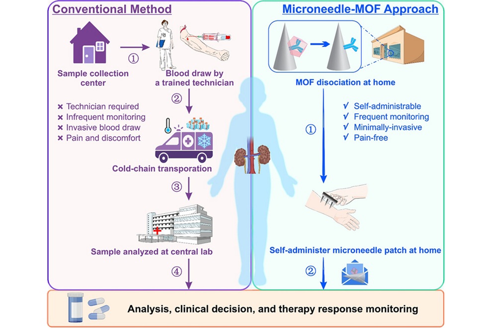
Microneedle Patch Enables At-Home Monitoring of Acute Kidney Injury
Kidney disease is often silent in its early stages, making timely detection difficult outside clinical settings. Early biomarkers such as neutrophil gelatinase–associated lipocalin can require blood draws... Read more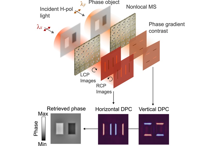
Ultrathin Metasurface Could Bring Quantitative Phase Imaging to Portable Devices
Point-of-care imaging often depends on bulky optical systems that have difficulty visualizing nearly transparent cells and tissues without staining. These limitations can slow diagnosis and restrict use... Read more
Business
view channel
Robotic Single-Port System Gains CE Mark for Transvaginal Gynecologic Procedures
Intuitive (Sunnyvale, CA, USA) announced that it has received CE mark approval for use of the da Vinci SP Single Port surgical system in transvaginal gynecologic procedures, marking the first such indication... Read more