Novel Surgical Procedure Treats Canalicular Obstruction
|
By HospiMedica International staff writers Posted on 27 Oct 2021 |
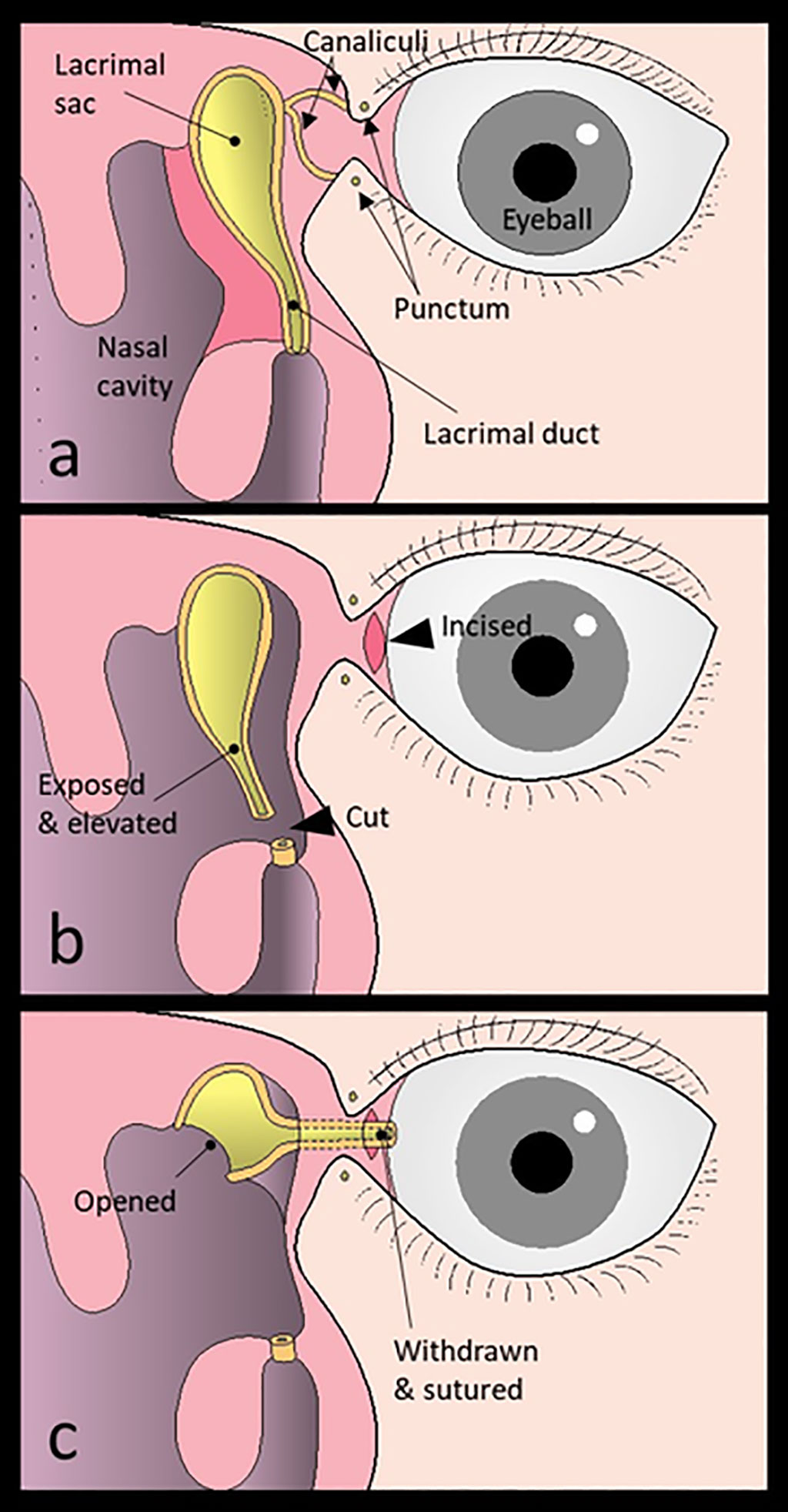
Image: Novel Surgical Procedure Treats Canalicular Obstruction (Photo courtesy of Munetaka Ushio/ Toho University)
A new surgical procedure overcomes the disadvantages of conventional methods to treat epiphora resulting from intractable obstruction of the tear ducts.
The new procedure, developed at Toho University (Tokyo, Japan), is called conjunctivoductivo-dacryocystorhinostomy, and is designed to treat watery eyes (epiphora), which affects the quality of life of those suffering from it. Performed under general anesthesia (GA), the procedure includes exposure and elevation of the entire lacrimal duct and lacrimal sac. The lacrimal duct is then cut at the distal end, and the conjunctiva is incised at the medial canthus. The cut end of the lacrimal duct is withdrawn from the conjunctival incision and sutured to form a new lacrimal punctum.
Subsequently, the medial wall of the lacrimal sac is opened, resulting in the former lacrimal duct and lacrimal sac becoming the new lacrimal passage, allowing tear fluids to flow through the newly made passage. The new procedure can be used to enlarge the canaliculus whenever a Jones tube--a semi-permanent glass tube that connects the nasal cavity and medical canthus--or external surgery are being considered. Both of these options leave a scar of approximately two cm on the side of the nose. The study was published on October 1, 2021 in The Laryngoscope.
“The newly developed procedure, 'conjunctivoductivo-dacryocystorhinostomy' for the intractable canalicular obstruction does not leave any facial scarring or place foreign matter in the body. We believe that this procedure can help improve the quality of life of patients with epiphora,” concluded lead author Munetaka Ushio, MD, PhD, and colleagues of the departments of ophthalmology and otolaryngology.
Under normal conditions, tears in the eyes are drained into the lacrimal sac through the upper and lower puncta, which are located in medial canthus. The tears drain through the superior and inferior canaliculi and onto the lacrimal sac and the nasolacrimal duct, and finally into the nose. When both upper and lower canaliculi are obstructed, tears cannot drain into the nose, resulting in epiphora.
Related Links:
Toho University
Latest Surgical Techniques News
- Minimally Invasive Endoscopic Surgery Improves Severe Stroke Outcomes
- Novel Glue Prevents Complications After Breast Cancer Surgery
- Breakthrough Brain Implant Enables Safer and More Precise Drug Delivery
- Bioadhesive Sponge Stops Uncontrolled Internal Bleeding During Surgery
- Revolutionary Nano Bone Material to Accelerate Surgery and Healing
- Superior Orthopedic Implants Combat Infections and Quicken Healing After Surgery
- Laser-Based Technique Eliminates Pancreatic Tumors While Protecting Healthy Tissue
- Surgical Treatment of Severe Carotid Artery Stenosis Benefits Blood-Brain Barrier
- Revolutionary Reusable Duodenoscope Introduces 68-Minute Sterilization
- World's First Transcatheter Smart Implant Monitors and Treats Congestion in Heart Failure
- Hybrid Endoscope Marks Breakthrough in Surgical Visualization
- Robot-Assisted Bronchoscope Diagnoses Tiniest and Hardest to Reach Lung Tumors
- Diamond-Titanium Device Paves Way for Smart Implants that Warn of Disease Progression
- 3D Printable Bio-Active Glass Could Serve as Bone Replacement Material
- Spider-Inspired Magnetic Soft Robots to Perform Minimally Invasive GI Tract Procedures
- Micro Imaging Device Paired with Endoscope Spots Cancers at Earlier Stage
Channels
Critical Care
view channel
AI Heart Attack Risk Assessment Tool Outperforms Existing Methods
For decades, doctors have relied on standardized scoring systems to assess patients with the most common type of heart attack—non-ST-elevation acute coronary syndrome (NSTE-ACS). The GRACE score, used... Read more
'Universal' Kidney to Match Any Blood Type
Blood-type incompatibility has long been one of the greatest obstacles in organ transplantation, forcing thousands of patients—particularly those with type O blood—to wait years longer for compatible donors.... Read morePatient Care
view channel
Revolutionary Automatic IV-Line Flushing Device to Enhance Infusion Care
More than 80% of in-hospital patients receive intravenous (IV) therapy. Every dose of IV medicine delivered in a small volume (<250 mL) infusion bag should be followed by subsequent flushing to ensure... Read more
VR Training Tool Combats Contamination of Portable Medical Equipment
Healthcare-associated infections (HAIs) impact one in every 31 patients, cause nearly 100,000 deaths each year, and cost USD 28.4 billion in direct medical expenses. Notably, up to 75% of these infections... Read more
Portable Biosensor Platform to Reduce Hospital-Acquired Infections
Approximately 4 million patients in the European Union acquire healthcare-associated infections (HAIs) or nosocomial infections each year, with around 37,000 deaths directly resulting from these infections,... Read moreFirst-Of-Its-Kind Portable Germicidal Light Technology Disinfects High-Touch Clinical Surfaces in Seconds
Reducing healthcare-acquired infections (HAIs) remains a pressing issue within global healthcare systems. In the United States alone, 1.7 million patients contract HAIs annually, leading to approximately... Read moreHealth IT
view channel
Printable Molecule-Selective Nanoparticles Enable Mass Production of Wearable Biosensors
The future of medicine is likely to focus on the personalization of healthcare—understanding exactly what an individual requires and delivering the appropriate combination of nutrients, metabolites, and... Read moreBusiness
view channel
Philips and Masimo Partner to Advance Patient Monitoring Measurement Technologies
Royal Philips (Amsterdam, Netherlands) and Masimo (Irvine, California, USA) have renewed their multi-year strategic collaboration, combining Philips’ expertise in patient monitoring with Masimo’s noninvasive... Read more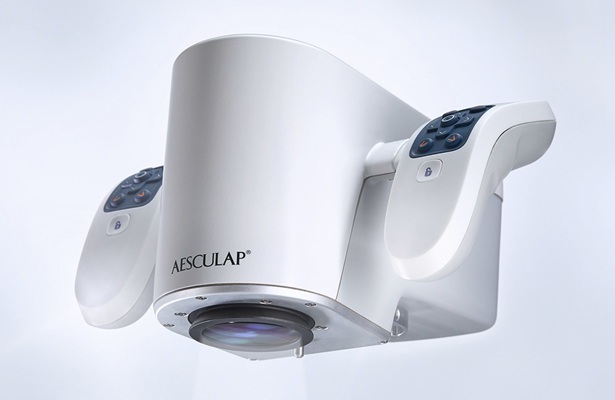
B. Braun Acquires Digital Microsurgery Company True Digital Surgery
The high-end microsurgery market in neurosurgery, spine, and ENT is undergoing a significant transformation. Traditional analog microscopes are giving way to digital exoscopes, which provide improved visualization,... Read more
CMEF 2025 to Promote Holistic and High-Quality Development of Medical and Health Industry
The 92nd China International Medical Equipment Fair (CMEF 2025) Autumn Exhibition is scheduled to be held from September 26 to 29 at the China Import and Export Fair Complex (Canton Fair Complex) in Guangzhou.... Read more


