Bioprinting of Bone Constructs with Genes During Surgery to Facilitate Long-Term Healing
|
By HospiMedica International staff writers Posted on 22 Apr 2022 |
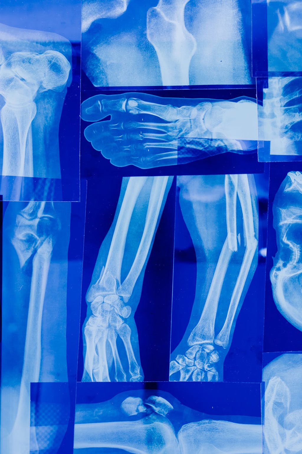
Given enough time and energy, the body will heal, but when doctors or engineers intervene, the processes do not always proceed as planned because chemicals that control and facilitate the healing process are missing. Now, an international team of engineers is bioprinting bone along with two growth factor encoding genes that help incorporate the cells and heal defects in the skulls of rats.
Researchers at Penn State (University Park, PA) used gene encoding PDGF-B, platelet derived-growth factor, which encourages cells to multiply and to migrate, and gene encoding BMP-2, bone morphogenetic protein, which improves bone regeneration. They delivered both genes using bioprinting. The team embedded the DNA for the protein in plasmids – ring-like loops of DNA that can transport genetic information. Once the DNA enters the progenitor cell, it begins to produce the appropriate proteins to enhance bone growth.
The two genes were printed during surgery onto a hole in the skull of a rat using a device very similar to an ink-jet printer. The mixture was created to release a burst of PDGF-B encoding gene in 10 days and a continuing release of BMP-2 encoding gene for five weeks. The rats that received bioprinted genes with controlled release of BMP-2 encoding gene saw about 40% bone tissue creation and 90% bone coverage in six weeks compared to 10% new bone tissue and 25% bone coverage for rats with the same defect, but no treatment.
"Growth factors are essential for cell growth. We use two different genes encoding two different growth factors. These growth factors help stem cells to migrate into the defect area and then help the progenitor cells to convert into bone," said Ibrahim T. Ozbolat, associate professor of engineering science and mechanics. "This method is better than simply dumping the growth factors. If we do that, the amounts of proteins are finite, but if we use gene therapy, the cells continue to produce the necessary growth factors."
Related Links:
Penn State
Latest Surgical Techniques News
- Robotic Assistant Delivers Ultra-Precision Injections with Rapid Setup Times
- Minimally Invasive Endoscopic Surgery Improves Severe Stroke Outcomes
- Novel Glue Prevents Complications After Breast Cancer Surgery
- Breakthrough Brain Implant Enables Safer and More Precise Drug Delivery
- Bioadhesive Sponge Stops Uncontrolled Internal Bleeding During Surgery
- Revolutionary Nano Bone Material to Accelerate Surgery and Healing
- Superior Orthopedic Implants Combat Infections and Quicken Healing After Surgery
- Laser-Based Technique Eliminates Pancreatic Tumors While Protecting Healthy Tissue
- Surgical Treatment of Severe Carotid Artery Stenosis Benefits Blood-Brain Barrier
- Revolutionary Reusable Duodenoscope Introduces 68-Minute Sterilization
- World's First Transcatheter Smart Implant Monitors and Treats Congestion in Heart Failure
- Hybrid Endoscope Marks Breakthrough in Surgical Visualization
- Robot-Assisted Bronchoscope Diagnoses Tiniest and Hardest to Reach Lung Tumors
- Diamond-Titanium Device Paves Way for Smart Implants that Warn of Disease Progression
- 3D Printable Bio-Active Glass Could Serve as Bone Replacement Material
- Spider-Inspired Magnetic Soft Robots to Perform Minimally Invasive GI Tract Procedures
Channels
Critical Care
view channel
CPR Guidelines Updated for Pediatric and Neonatal Emergency Care and Resuscitation
Cardiac arrest in infants and children remains a leading cause of pediatric emergencies, with more than 7,000 out-of-hospital and 20,000 in-hospital cardiac arrests occurring annually in the United States.... Read more
Ingestible Capsule Monitors Intestinal Inflammation
Acute mesenteric ischemia—a life-threatening condition caused by blocked blood flow to the intestines—remains difficult to diagnose early because its symptoms often mimic common digestive problems.... Read more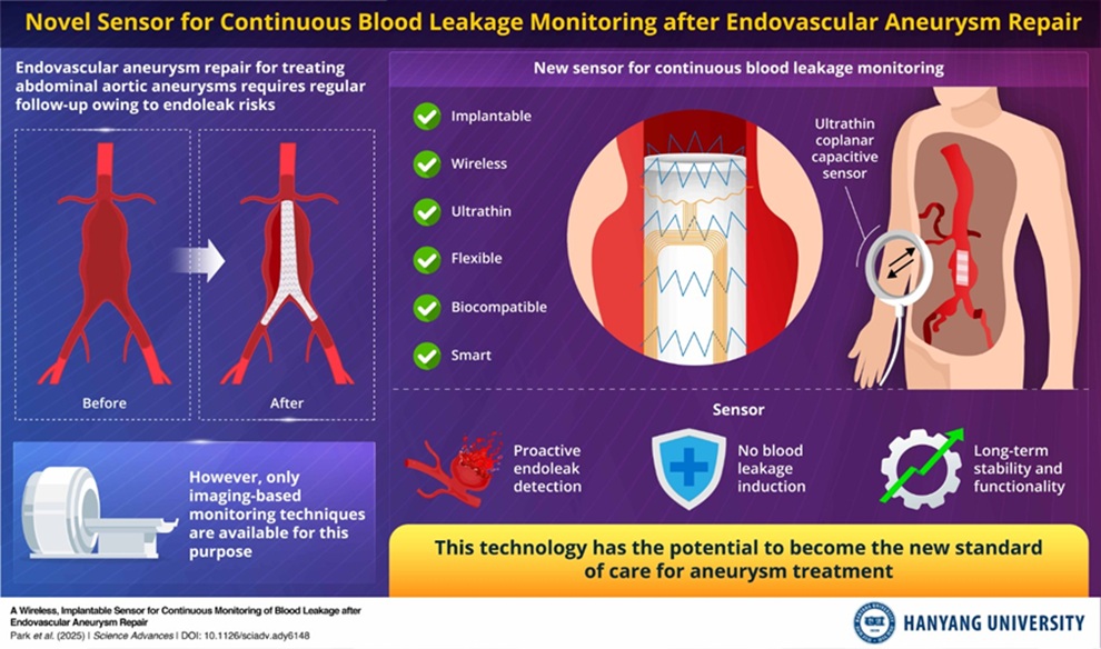
Wireless Implantable Sensor Enables Continuous Endoleak Monitoring
Endovascular aneurysm repair (EVAR) is a life-saving, minimally invasive treatment for abdominal aortic aneurysms—balloon-like bulges in the aorta that can rupture with fatal consequences.... Read more
Wearable Patch for Early Skin Cancer Detection to Reduce Unnecessary Biopsies
Skin cancer remains one of the most dangerous and common cancers worldwide, with early detection crucial for improving survival rates. Traditional diagnostic methods—visual inspections, imaging, and biopsies—can... Read morePatient Care
view channel
Revolutionary Automatic IV-Line Flushing Device to Enhance Infusion Care
More than 80% of in-hospital patients receive intravenous (IV) therapy. Every dose of IV medicine delivered in a small volume (<250 mL) infusion bag should be followed by subsequent flushing to ensure... Read more
VR Training Tool Combats Contamination of Portable Medical Equipment
Healthcare-associated infections (HAIs) impact one in every 31 patients, cause nearly 100,000 deaths each year, and cost USD 28.4 billion in direct medical expenses. Notably, up to 75% of these infections... Read more
Portable Biosensor Platform to Reduce Hospital-Acquired Infections
Approximately 4 million patients in the European Union acquire healthcare-associated infections (HAIs) or nosocomial infections each year, with around 37,000 deaths directly resulting from these infections,... Read moreFirst-Of-Its-Kind Portable Germicidal Light Technology Disinfects High-Touch Clinical Surfaces in Seconds
Reducing healthcare-acquired infections (HAIs) remains a pressing issue within global healthcare systems. In the United States alone, 1.7 million patients contract HAIs annually, leading to approximately... Read moreHealth IT
view channel
Printable Molecule-Selective Nanoparticles Enable Mass Production of Wearable Biosensors
The future of medicine is likely to focus on the personalization of healthcare—understanding exactly what an individual requires and delivering the appropriate combination of nutrients, metabolites, and... Read moreBusiness
view channel
Philips and Masimo Partner to Advance Patient Monitoring Measurement Technologies
Royal Philips (Amsterdam, Netherlands) and Masimo (Irvine, California, USA) have renewed their multi-year strategic collaboration, combining Philips’ expertise in patient monitoring with Masimo’s noninvasive... Read more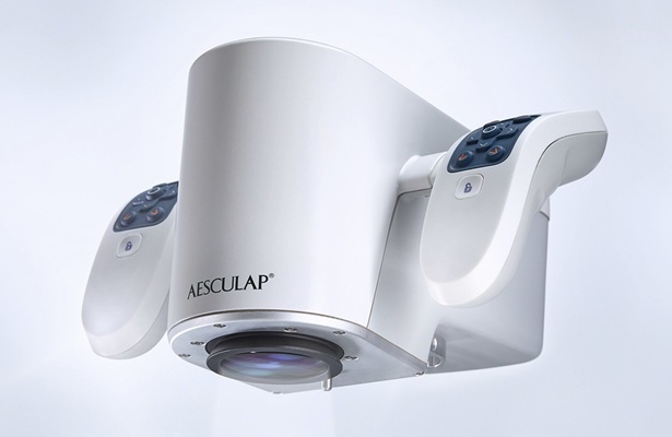
B. Braun Acquires Digital Microsurgery Company True Digital Surgery
The high-end microsurgery market in neurosurgery, spine, and ENT is undergoing a significant transformation. Traditional analog microscopes are giving way to digital exoscopes, which provide improved visualization,... Read more
CMEF 2025 to Promote Holistic and High-Quality Development of Medical and Health Industry
The 92nd China International Medical Equipment Fair (CMEF 2025) Autumn Exhibition is scheduled to be held from September 26 to 29 at the China Import and Export Fair Complex (Canton Fair Complex) in Guangzhou.... Read more