


New Coronary IVL Catheter Treats Severely Calcified Coronary Artery Disease Using Pulsatile Sonic Pressure
|
By HospiMedica International staff writers Posted on 24 May 2023 |


|
One in three patients with coronary artery disease have calcified lesions, with heavily calcified coronary lesions posing significant challenges in current coronary interventions due to difficulty in dilation. Existing treatment solutions, including high pressure and modified balloons, as well as atherectomy, face limitations due to their inability to reliably dilate vessels with rigid, severe calcium. These technologies only treat superficial calcium, leaving deep calcium untreated, which can affect procedural outcomes.
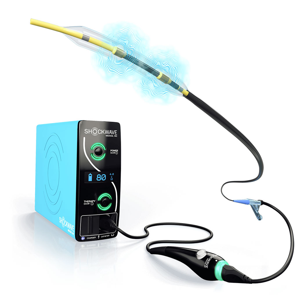
Now, the latest device to treat severely calcified coronary artery disease - Shockwave Intravascular Lithotripsy (S-IVL) System with the Shockwave C2 Coronary Intravascular Lithotripsy (IVL) Catheter from Shockwave Medical (Santa Clara, CA, USA) – effectively addresses these challenges. IVL utilizes sonic pressure waves to break down calcium in situ. After inflating the integrated balloon to 4-atm, a small spark at the emitters vaporizes the saline-contrast solution and generates a bubble. This bubble quickly expands and collapses within the balloon, resulting in a brief burst of sonic pressure waves. These waves pass through the coronary tissue and reflect off the calcium, breaking it down with an effective pressure of about 50 atm. The emitters positioned along the length of the device create a localized field within the vessel to fracture both intimal and medial calcium. The integrated balloon plays a crucial role; it positions against the vessel wall to facilitate efficient energy transfer during IVL, then helps to dilate the lesion to maximize lumen gain.
The Shockwave C2+ Coronary IVL Catheter is specifically designed to treat longer calcified lesions and more challenging eccentric and nodular calcium. It minimizes trauma to soft tissue by safely targeting and fracturing intimal and medial calcium. This optimizes stent delivery, expansion and apposition while reducing the risk of perforation and cost escalation. With its straightforward and intuitive system, it makes complex calcified coronary procedures more predictable. The Shockwave C2+ offers 50% more pulses per catheter than Shockwave C2, making it particularly suitable for treating longer calcified lesions and more challenging eccentric and nodular calcium.
“Shockwave C2+ maintains the intuitive catheter design and ease of use that are foundational to the success of Shockwave IVL and incorporates improvements that will enhance procedural efficiency and optimize the treatment of the most challenging morphologies,” said Jonathan Hill, MD, Consultant Cardiologist at London’s Royal Brompton Hospital. “The extra pulses are most advantageous in areas with the highest burden of calcium, including nodular, eccentric, diffuse and multivessel calcium.”
Related Links:
Shockwave Medical




Latest Critical Care News
- AI-Derived Heart Age Linked to Cardiovascular and Dementia-Related Mortality
- Updated Head Injury Guidelines Support Risk-Based CT and Observation Decisions
- Inhaled Therapy Uses Modified Bacteria to Break Down Pseudomonas Biofilms
- Miniature Wearable Sensor Enables Continuous Cuffless Blood Pressure Monitoring
- Studies Support AI-ECG for Detecting Suspected Hypertrophic Cardiomyopathy
- Updated Staging System Standardizes Cardiogenic Shock Assessment
- Moldable Sensor Could Support Neck Muscle Monitoring and Rehabilitation
- Routine Opt-Out Screening Helps Increase Detection of Adolescent STIs
- Personalized fMRI-Guided TMS May Improve Severe OCD Symptoms
- Ultrasound Implant Could Help Monitor Spinal Cord Recovery After Surgery
- Paper-Based Patch Integrates Sweat Sensors and Drug-Delivery Microneedles
- Circuit-Free Smart Patch Uses Smartphone Camera to Monitor Pulse and Vessel Stiffness

- Chest Digital Twin Guides Light Therapy Dosing for Pneumonia
- Intraoperative Drug Combination Reduces Kidney Injury in Septic Shock Surgery
- Wirelessly Powered Implant Targets Excess Glucose in the Blood
- Noninvasive Brain-Computer Interface Enables Responses in Some Unresponsive ICU Patients
Channels
Artificial Intelligence
view channel
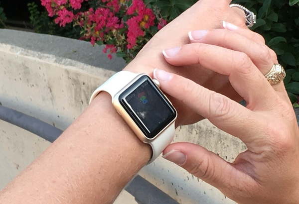
AI Analysis of Smartwatch ECGs May Help Detect Weakened Heart Function
Left ventricular systolic dysfunction (LVSD) occurs when the heart’s main pumping chamber does not contract effectively. The condition can remain asymptomatic for some time but may progress to symptomatic... Read more
AI Tool Speeds Echocardiogram Reporting With Comparable Accuracy
Echocardiography is the most widely used form of cardiac imaging, playing a key role in diagnosing heart disease and guiding treatment decisions. However, reviewing ultrasound images and documenting cardiac... Read more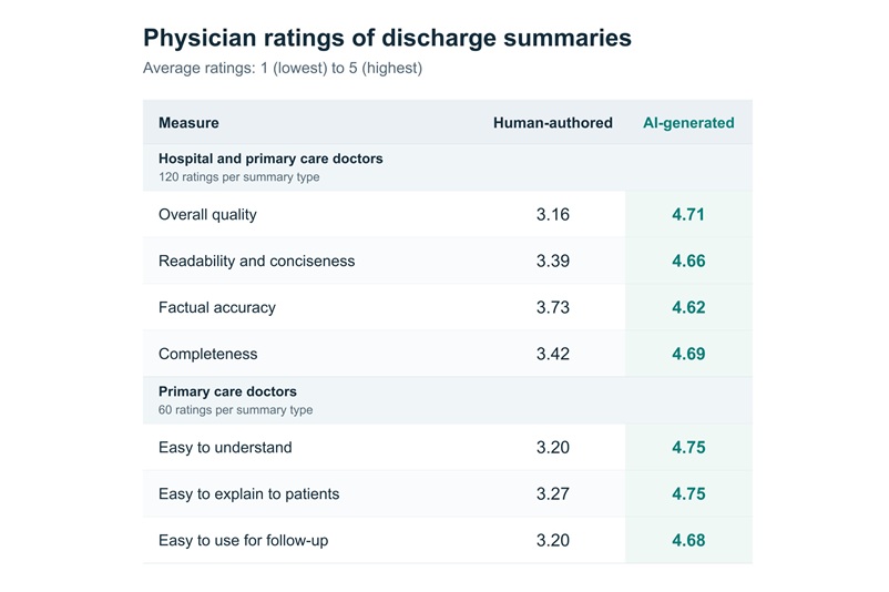
Surgical Techniques
view channel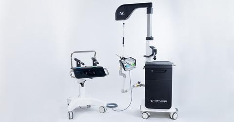
FDA Authorizes Robotic System for En Bloc Bladder Resection
Bladder cancer frequently recurs, making accurate staging essential for treatment planning. Conventional transurethral resection can fragment tumors, complicating margin assessment and detrusor muscle... Read more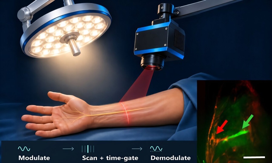
Fluorescence Imaging Reveals Nerves Under Normal Operating-Room Lighting
Preventing unintended nerve injury remains a major concern during surgery, particularly in complex anatomical regions where nerves can be difficult to distinguish from surrounding tissue.... Read more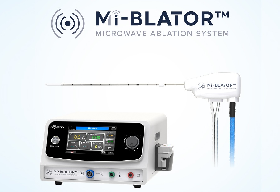
Programmable Microwave Ablation System Gains CE Mark for Minimally Invasive Tissue Ablation
Hospitals continue to seek minimally invasive approaches that simplify procedural planning and provide controlled energy delivery for tissue ablation. Thermal ablation systems are used across multiple... Read more
Minimally Invasive Balloon Procedure May Redirect Blood Flow During Cardiac Arrest
Out-of-hospital cardiac arrest has a low survival rate, and patients who do not respond to prolonged advanced resuscitation have few remaining treatment options. During cardiopulmonary resuscitation, blood... Read morePatient Care
view channel
Virtual Reality Program May Reduce Anxiety Before Pediatric Surgery
Presurgery anxiety affects an estimated 50% to 75% of children undergoing surgery and can complicate anesthesia induction, increase postoperative pain, and prolong recovery. Sedation is often used to manage... Read more
Video-Based Support Helps Older Adults Clarify Kidney Disease Treatment Preferences
Advanced chronic kidney disease (CKD) involves progressive loss of kidney function that can lead to kidney failure and difficult treatment decisions. For older adults, these decisions may include choosing... Read more
Medical Imaging
view channel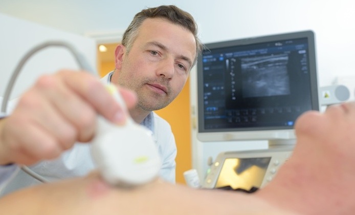
Lung Ultrasound Predicts Progression of Interstitial Lung Abnormalities in Rheumatoid Arthritis
Interstitial lung abnormalities are a pulmonary concern in patients with rheumatoid arthritis, requiring monitoring to detect disease progression. However, tracking these changes often involves repeated... Read more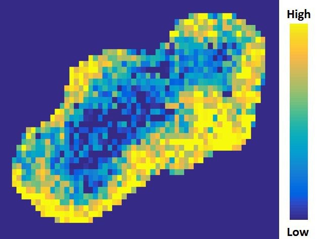
MRI Test Identifies Candidates for Lower-Dose Radiotherapy in HPV Throat Cancer
Human papillomavirus (HPV)-positive oropharyngeal cancer is a throat cancer linked to HPV infection. Its incidence is rising, and many patients receive high-dose radiotherapy that can cause long-term swallowing... Read more
Health IT
view channel
Clinical Decision Support Tool Automates CKD Risk Assessment in Primary Care
Chronic kidney disease (CKD) requires timely diagnosis, risk assessment, and coordinated treatment in primary care. Delayed recognition can affect decisions about testing, blood pressure control, cardiovascular... Read more
EHR Alerts Support Dosing, Sedation, and Shock Recognition in Cardiac Care
Electronic health record alerts can add to clinician workload when they are poorly targeted. In critical cardiac care, however, delayed decisions can affect treatment timing, sedation exposure, and escalation... Read more
Point of Care
view channel
Handheld Imaging Device May Help Identify Preterm Birth Risk Earlier
Preterm birth, defined as delivery before 37 weeks of pregnancy, can lead to serious neonatal complications, including neurological, developmental, and respiratory problems. However, identifying pregnancies... Read more
New Breath Condensate Testing Approach Enhances Reliability for COPD Monitoring
Chronic obstructive pulmonary disease (COPD) is a progressive lung condition that can increasingly impair breathing over time. Diagnosis and monitoring may be burdensome because patients often require... Read more
Continuous Glucose Monitoring Identifies Cardiometabolic Risk in Adults Without Diabetes
Dysglycemia—abnormal blood glucose regulation—can fluctuate throughout the day and often escape conventional screening. Clinicians typically rely on fasting plasma glucose and hemoglobin A1c, which offer... Read more
Business
view channel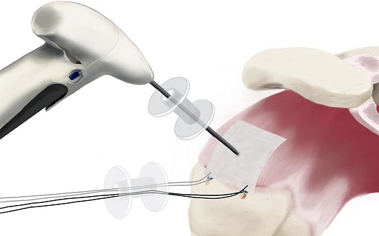
Stryker Completes Acquisition to Broaden Shoulder Surgery Offerings
Stryker (Portage, MI, USA) has completed its acquisition of ZuriMED (Zurich, Switzerland), a privately held developer of the FiberLocker System, expanding its Sports Medicine portfolio with a commercialized... Read more