



AI Tool Accurately Predicts Cancer Three Years Prior to Diagnosis
|
By HospiMedica International staff writers Posted on 24 Aug 2023 |


|

Over the past five decades, there has been a surge in cases of a specific type of esophageal and stomach cancer - esophageal adenocarcinoma (EAC) and gastric cardia adenocarcinoma (GCA). Both cancers have high fatality rates, although preventive measures can make a difference. Screenings can detect pre-cancerous changes, such as Barrett’s esophagus, often identified in individuals with long-standing gastroesophageal reflux disease (GERD). Although guidelines suggest screening for high-risk patients, many healthcare providers remain unfamiliar with this recommendation. Now, a new artificial intelligence (AI) tool offers accurate predictions for these forms of esophageal and stomach cancer at least three years in advance of a diagnosis.
Researchers at Michigan Medicine (Ann Arbor, MI, USA) have developed an automated tool integrated into the electronic health record (EHR). This tool has the potential to bridge the awareness gap between healthcare providers and patients with an elevated risk of developing EAC and GCA. The researchers employed a specific type of AI for analyzing data related to EAC and GCA rates across more than 10 million U.S. veterans in order to develop and validate the Kettles Esophageal and Cardia Adenocarcinoma prediction tool, or K-ECAN for short.
K-ECAN leverages readily accessible data from the EHR, including patient demographics, weight, prior diagnoses, and routine lab results, to gauge an individual's risk of developing EAC and GCA. Outperforming published guidelines and previously validated prediction tools, K-ECAN accurately predicts cancer at least three years ahead of a diagnosis. Integrating this AI tool into the EHR could automatically notify healthcare providers about patients at a higher risk of developing EAC and GCA.
“Symptoms of GERD, like heartburn, are an important risk factor for esophageal adenocarcinoma,” said Joel Rubenstein, M.D., M.S., professor of internal medicine at Michigan Medicine. “But most people with GERD symptoms will never develop esophageal adenocarcinoma and gastric cardia adenocarcinoma. In addition, roughly half of the patients with this form of cancer never experienced prior GERD symptoms at all. This makes K-ECAN particularly useful because it can identify people who are at elevated risk, regardless of whether they have GERD symptoms or not.”
Related Links:
Michigan Medicine






Latest AI News
- Optical Imaging System Helps Predict Chemotherapy Response in Cancer
- AI Tool Predicts Long-Term Cardiovascular Risk from Sleep Study ECG
- Predictive Model Supports Growth Hormone Treatment Planning in Idiopathic Short Stature
- AI Model Predicts Prolonged Sitting in Chronic Pelvic Pain
- Autonomous AI Helps Streamline Urgent Skin Cancer Referrals
- Virtual Ultrasound Images Support Development of Cardiovascular Imaging Tools
- Explainable AI Tool Improves Brain Disorder Screening from 4D fMRI
- New Benchmark Highlights Evidence Gaps in AI Responses to Clinical Questions
- AI-Powered ECG Model Gains FDA Authorization for Emergency Cardiac Assessment
- AI Improves Non-Contrast CT Interpretation for Time-Sensitive Stroke Assessment
- Explainable AI Tool Identifies High-Risk Patients Before Heart Attack Reperfusion
- AI Tools Show Potential to Detect Subtle Signs of Interval Breast Cancer
- AI Framework Aims to Turn Medical Care from Reactive to Preventive
Channels
Critical Care
view channel
Proposed Risk Score Prioritizes Sickest Patients for Heart Transplant
Donor hearts remain extremely scarce in the U.S. transplant system, with one in five candidates dying or being removed from the waiting list before receiving an organ. Current allocation rules may also... Read more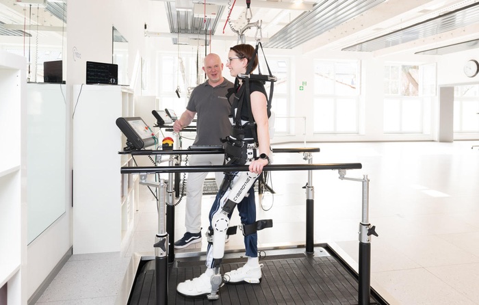
Wearable Robotic Gait Training Linked to Improved Walking Distance in ALS
Amyotrophic lateral sclerosis (ALS) is a progressive neuromuscular disease that damages nerve cells controlling voluntary muscles. As the disease advances, patients can lose the ability to walk, speak,... Read more
Surgical Techniques
view channel
New Surgical Technique Enlarges Aortic Root for Larger Valve Implantation
When replacing a diseased aortic valve, a small aortic root can prevent surgeons from implanting a prosthesis large enough for the patient’s body size. This can lead to patient-prosthesis mismatch, causing... Read more
FDA Approval Expands Bridging Stent Use in Complex Aortic Aneurysm Repair
Complex aortic aneurysms are increasingly treated with fenestrated and branched endovascular aortic repair, which rely on bridging stents to maintain blood flow to visceral arteries. These complex procedures... Read more
Patient Care
view channel
AI Platform Personalizes Support Between Visits for Cancer Survivors
Cancer survivors often manage persistent side effects after treatment, including fatigue, anxiety, uncertainty, symptoms, stress, and questions between appointments. Clinical teams need ways to maintain... Read more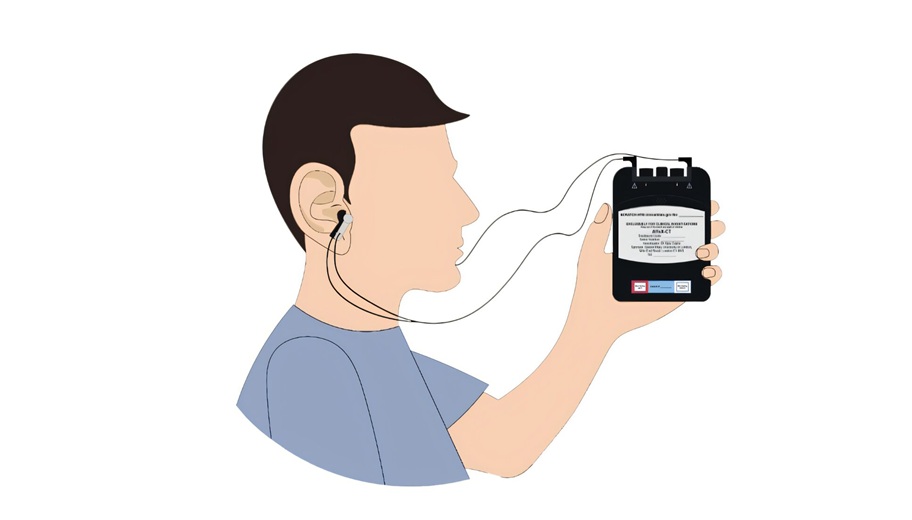
At-Home Neuromodulation Shows Promise in Selected Patients With Hypertension
High blood pressure is a major risk factor for cardiovascular disease, including heart attack and stroke. Many patients receiving treatment for hypertension still remain above recommended blood pressure... Read more
EHR Model Flags High-Risk Periods in Patients with Metastatic Breast Cancer
Determining when patients with metastatic breast cancer are nearing the end of life remains difficult, often leading to intensive interventions with limited benefit. Prognostic uncertainty can delay goals-of-care... Read more
Medical Imaging
view channel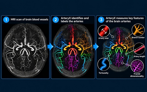
MRI Tool Analysis Identifies Vascular Differences in Cerebral Small Vessel Disease
Cerebral small vessel disease (CSVD) involves changes or damage to the brain’s small blood vessels and is associated with stroke and cognitive impairment. Magnetic resonance imaging (MRI) can show cerebral... Read more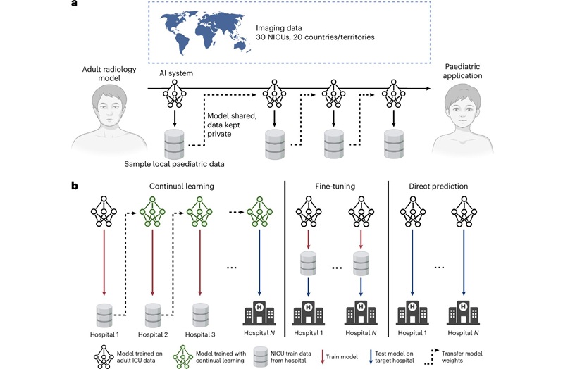
Privacy-Preserving Learning Model Adapts Adult Radiology AI for Neonatal Care
Artificial intelligence models may support radiology image interpretation, yet pediatric and neonatal care remain underserved. Privacy restrictions limit access to children’s medical records, and pediatric... Read more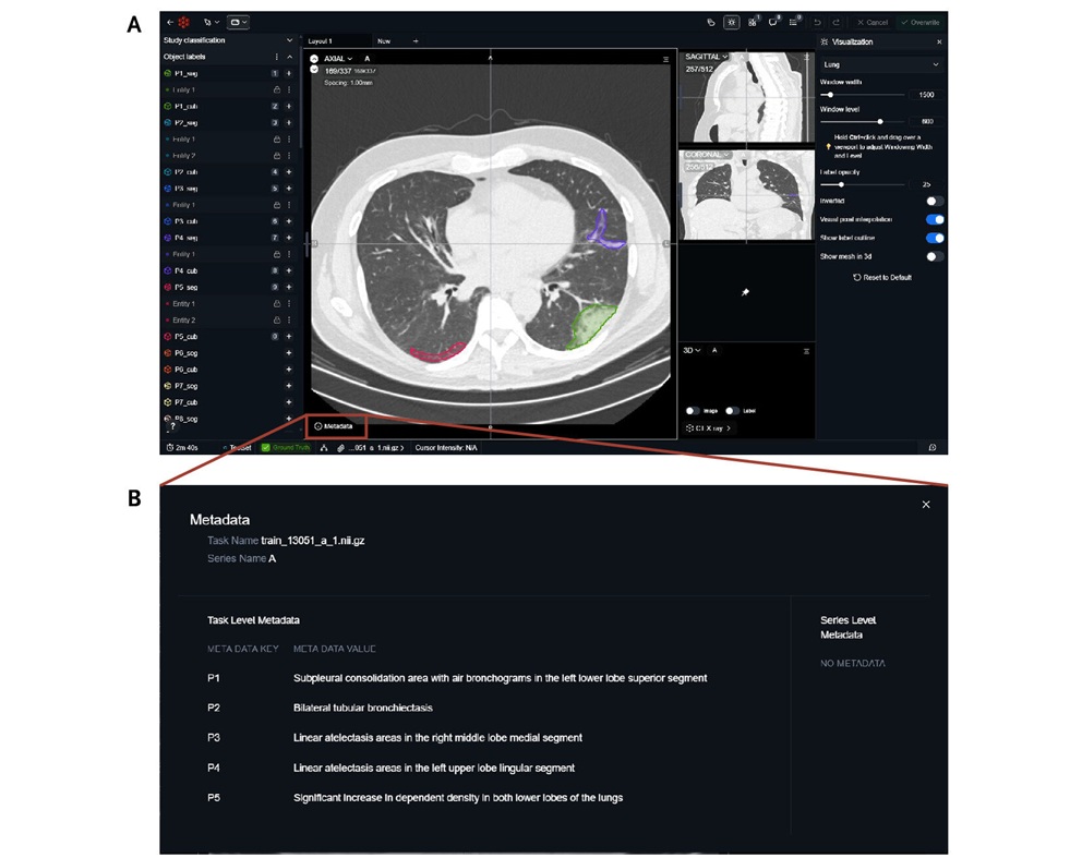
Public Chest CT Dataset Supports Explainable AI in Diagnostic Radiology
Chest computed tomography scans generate complex three-dimensional images that can be difficult for artificial intelligence systems to explain clearly. Clinicians need to verify AI findings before using... Read more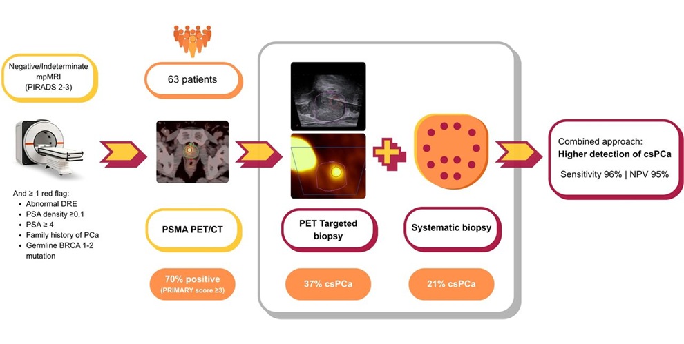
PSMA PET/CT-Guided Biopsy Improves Detection of Significant Prostate Cancer After Nondiagnostic MRI
Prostate cancer can be difficult to detect when magnetic resonance imaging (MRI) findings are negative or inconclusive. Men with persistent clinical risk factors may still require biopsy, yet standard... Read moreHealth IT
view channel
EHR Alerts Support Dosing, Sedation, and Shock Recognition in Cardiac Care
Electronic health record alerts can add to clinician workload when they are poorly targeted. In critical cardiac care, however, delayed decisions can affect treatment timing, sedation exposure, and escalation... Read more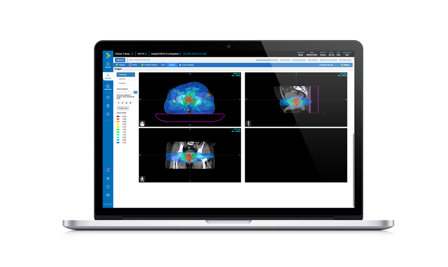
Adaptive Radiation QA Software Gains Validation for NeoArc Treatments
Adaptive radiation therapy allows clinicians to adjust treatment plans to daily anatomical changes such as tumor shrinkage, weight loss, or organ movement, improving precision while limiting exposure to... Read more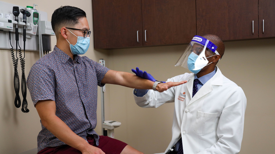

Point of Care
view channel
Continuous Glucose Monitoring Identifies Cardiometabolic Risk in Adults Without Diabetes
Dysglycemia—abnormal blood glucose regulation—can fluctuate throughout the day and often escape conventional screening. Clinicians typically rely on fasting plasma glucose and hemoglobin A1c, which offer... Read more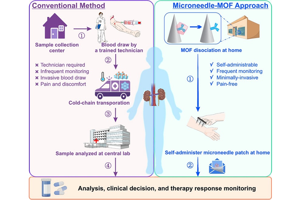
Microneedle Patch Enables At-Home Monitoring of Acute Kidney Injury
Kidney disease is often silent in its early stages, making timely detection difficult outside clinical settings. Early biomarkers such as neutrophil gelatinase–associated lipocalin can require blood draws... Read more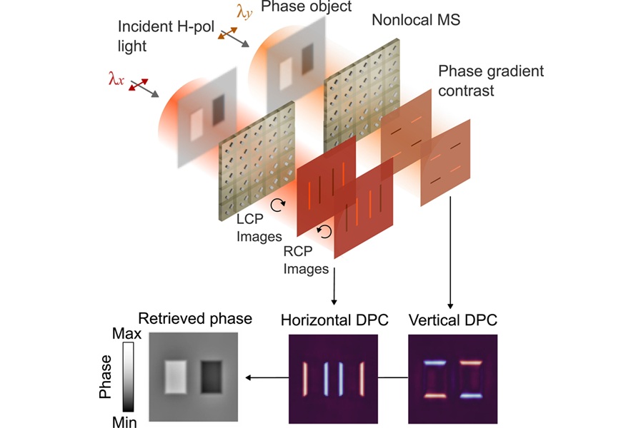
Business
view channel
Terumo Neuro Acquisition Advances Liquid Embolic Technology for Chronic Subdural Hematoma
Terumo Neuro (Aliso Viejo, CA, USA) signed an agreement to acquire Arsenal Medical Inc. (Waltham, MA, USA), a privately held, clinical-stage biomaterials company. The proposed acquisition would add NeoCast,... Read more
World Health Expo to Showcase Trillion-Dollar Growth Across Pharma, Biotech, Longevity and Wellness at 2027 Edition
World Health Expo (WHX) expands with the new Pharma, Biotech, Longevity and Wellness industry sectors More than 235,000 professional visits, over 4,200 exhibitors and representatives from over... Read more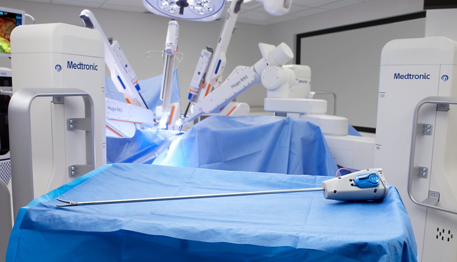
Medtronic Receives FDA Clearance for Vessel-Sealing Instrument for Robotic Surgery
As robotic-assisted surgery expands across U.S. hospitals, teams increasingly seek energy instruments with the familiarity and performance of tools used in open and laparoscopic procedures.... Read more