



Wireless, Handheld, Non-Invasive Device Detects Alzheimer's and Parkinson's Biomarkers
|
By HospiMedica International staff writers Posted on 21 Nov 2023 |


|
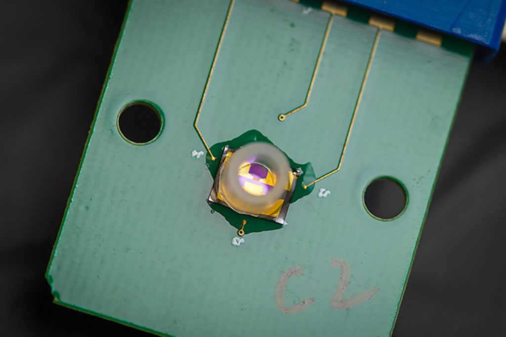
The prevalence of neurodegenerative diseases like Alzheimer’s and Parkinson’s is increasing. Current diagnostic methods for these conditions typically involve invasive procedures such as spinal taps and MRI scans, which can be daunting for patients and pose challenges for those with mobility issues or limited access to medical facilities. In response to these challenges, researchers have developed a groundbreaking handheld device that non-invasively detects biomarkers associated with Alzheimer’s and Parkinson’s Diseases. Additionally, this device has the capability to wirelessly transmit its findings to a computer or smartphone.
Developed by an international team of researchers led by UC San Diego (La Jolla, CA, USA), the device utilizes an electrical detection system rather than a chemical one. This approach is both simpler and more accurate. The core component of the device is a highly sensitive transistor, known as a field effect transistor (FET), featuring a graphene layer that is just one atom thick. This graphene FET (GFET) incorporates three electrodes: the source and drain electrodes connected to a battery’s positive and negative poles to facilitate current flow, and a gate electrode that regulates this current. A unique DNA strand, acting as a probe, is attached to the gate electrode. This probe specifically binds to amyloid beta, tau, or synuclein proteins. The interaction between these amyloids and their corresponding DNA probe alters the current between the source and drain electrodes, and it is this change in current or voltage that signals the presence of specific biomarkers.
The device’s effectiveness was validated using brain-derived amyloid proteins from deceased Alzheimer’s and Parkinson’s patients. The biosensors accurately detected the disease-specific biomarkers, matching the precision of current advanced diagnostic methods. Remarkably, the device requires only minimal sample quantities and can accurately perform even in samples containing a variety of proteins. While detecting tau proteins presented more challenges, the device’s ability to analyze multiple biomarkers allows for a comprehensive and reliable diagnosis. Future plans include testing the device with blood plasma, cerebrospinal fluid, and eventually saliva and urine samples in hospitals and nursing homes. Following successful trials, the researchers aim to seek FDA approval within the next few months, with the ultimate goal of commercializing the device within a year.
“This portable diagnostic system would allow testing at-home and at point of care, like clinics and nursing homes, for neurodegenerative diseases globally,” said Ratnesh Lal, a bioengineering, mechanical engineering and materials science professor at the UC San Diego Jacobs School of Engineering.
Related Links:
UC San Diego





Latest Point of Care News
- Continuous Glucose Monitoring Identifies Cardiometabolic Risk in Adults Without Diabetes
- Microneedle Patch Enables At-Home Monitoring of Acute Kidney Injury
- Ultrathin Metasurface Could Bring Quantitative Phase Imaging to Portable Devices
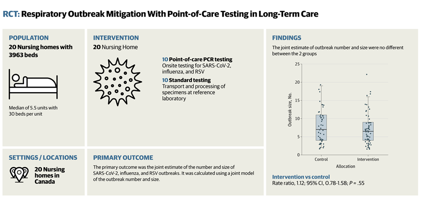
- Rapid Onsite Testing Reduces Emergency Department Transfers in Long-Term Care
- New Brain Ultrasound Platform Enables Bedside Postoperative Imaging
- Handheld Ultrasound Expands Point-of-Care Imaging Access in Brazil
- AI Dermatology Platform Targets Early Detection of Non-Melanoma Skin Cancer
- Handheld AI Device for Point-of-Care Skin Lesion Assessment Receives CE Mark
- Portable Immunoassay System Advances Toward Point-of-Care Biomarker Testing
- Portable MRI System Accelerates Emergency Brain Imaging and Triage
- Point-of-Care Viscoelastic Testing System Supports Obstetric Bleeding Management
- Portable MRI System Gains CE Mark for Point-of-Care Brain Imaging
Channels
Artificial Intelligence
view channel
AI Tool Predicts Cancer Survival and Biological Age from Single Facial Photo
Assessing frailty and physiologic reserve remains challenging when clinicians consider intensive cancer treatment. Chronological age alone may not reflect how well a patient can tolerate radiation, surgery,... Read more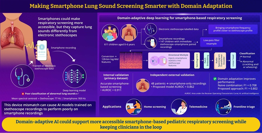
AI Tool Improves Smartphone Detection of Abnormal Pediatric Lung Sounds
Respiratory disease in children remains a major clinical concern worldwide. Abnormal lung sounds can indicate pediatric respiratory illness, but standard auscultation usually requires an in-person examination... Read more

Critical Care
view channel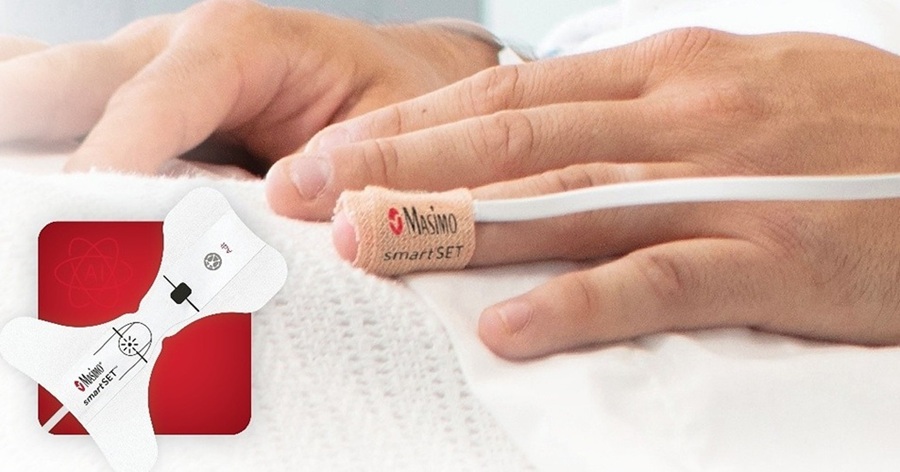
Smart Pulse Oximetry Supports Earlier Recognition of Arrhythmias and Respiratory Risk
On general care floors, respiratory compromise and arrhythmias can develop between routine checks. Atrial fibrillation often goes undiagnosed in older adults, while opioid-induced respiratory depression... Read more
Proposed Risk Score Prioritizes Sickest Patients for Heart Transplant
Donor hearts remain extremely scarce in the U.S. transplant system, with one in five candidates dying or being removed from the waiting list before receiving an organ. Current allocation rules may also... Read more

Surgical Techniques
view channel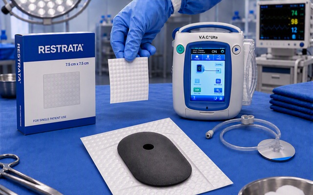
Combined Wound Healing Approach Pairs Synthetic Scaffold With Negative Pressure Therapy
Managing complex wounds requires both structural support for tissue repair and a controlled environment that protects the healing process. Frequent dressing changes can disrupt the wound bed and add complexity... Read more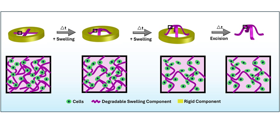
Shape-Changing Hydrogels Show Promise for Tissue Engineering and Wound Healing
Human tissues often have complex three-dimensional shapes that are closely tied to their function, but recreating these curvatures remains challenging in tissue engineering and wound repair.... Read more
Patient Care
view channel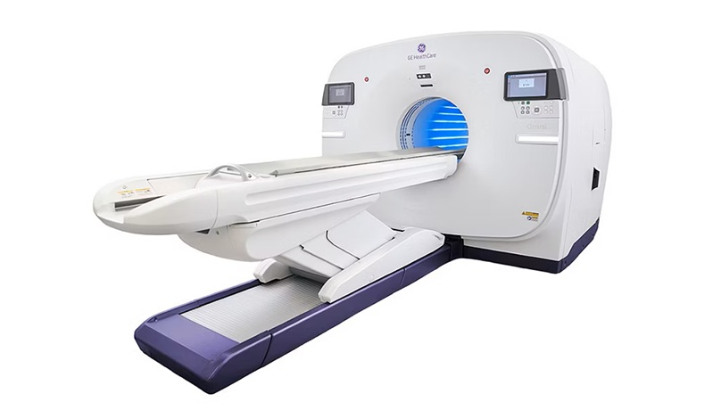
Total-Body PET/CT Scanner Enables One-Minute Diagnostic-Quality Cancer Imaging
Positron emission tomography/computed tomography (PET/CT) combines radiotracer-based functional imaging with anatomical detail to diagnose, stage, and monitor cancer. Conventional scanners image the body... Read more
AI Platform Personalizes Support Between Visits for Cancer Survivors
Cancer survivors often manage persistent side effects after treatment, including fatigue, anxiety, uncertainty, symptoms, stress, and questions between appointments. Clinical teams need ways to maintain... Read more
At-Home Neuromodulation Shows Promise in Selected Patients With Hypertension
High blood pressure is a major risk factor for cardiovascular disease, including heart attack and stroke. Many patients receiving treatment for hypertension still remain above recommended blood pressure... Read more
EHR Model Flags High-Risk Periods in Patients with Metastatic Breast Cancer
Determining when patients with metastatic breast cancer are nearing the end of life remains difficult, often leading to intensive interventions with limited benefit. Prognostic uncertainty can delay goals-of-care... Read moreMedical Imaging
view channel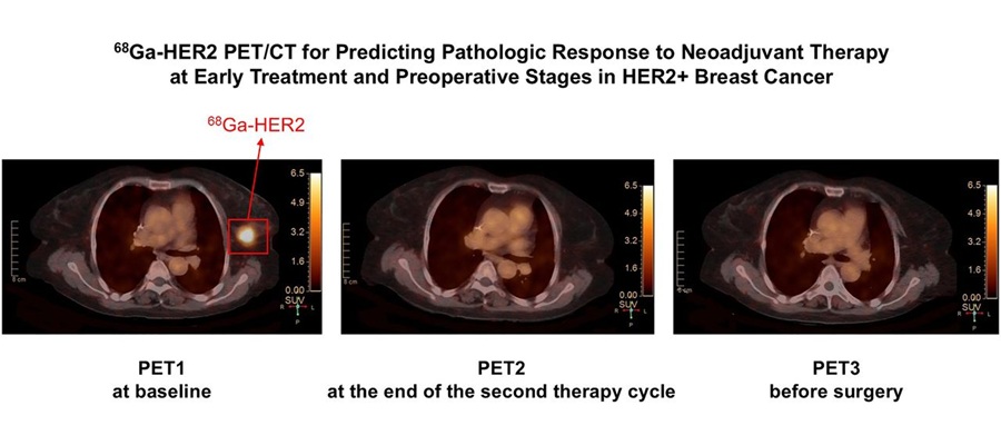
Targeted PET/CT Predicts Early Treatment Response in HER2-Positive Breast Cancer
HER2-positive breast cancer is an aggressive subtype that accounts for a substantial proportion of breast cancer cases. Patients often receive neoadjuvant therapy before surgery to shrink tumors, but assessing... Read more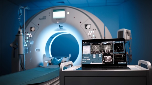
AI Model Detects Hidden Disease Signals in Routine CT Scans
Computed tomography (CT) scans are often performed to answer a specific diagnostic question, which can leave other potentially important findings outside the immediate clinical focus. As a result, opportunities... Read more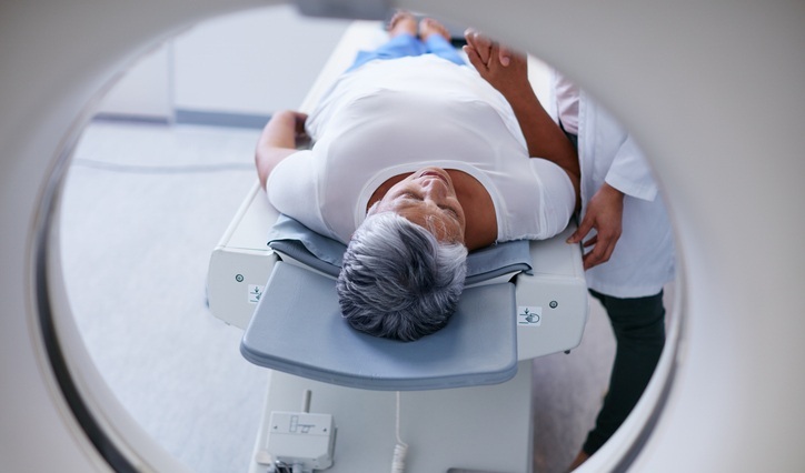
Amyloid and Tau PET Guide Selection and Monitoring of Alzheimer’s Therapy
Alzheimer’s disease is the most common form of dementia and can be difficult to distinguish from other causes of cognitive decline. According to the World Health Organization (WHO), 57 million people were... Read more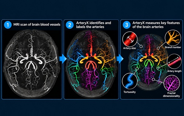
MRI Tool Analysis Identifies Vascular Differences in Cerebral Small Vessel Disease
Cerebral small vessel disease (CSVD) involves changes or damage to the brain’s small blood vessels and is associated with stroke and cognitive impairment. Magnetic resonance imaging (MRI) can show cerebral... Read moreHealth IT
view channel
EHR Alerts Support Dosing, Sedation, and Shock Recognition in Cardiac Care
Electronic health record alerts can add to clinician workload when they are poorly targeted. In critical cardiac care, however, delayed decisions can affect treatment timing, sedation exposure, and escalation... Read more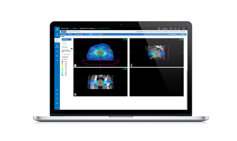
Adaptive Radiation QA Software Gains Validation for NeoArc Treatments
Adaptive radiation therapy allows clinicians to adjust treatment plans to daily anatomical changes such as tumor shrinkage, weight loss, or organ movement, improving precision while limiting exposure to... Read more

Point of Care
view channel
Continuous Glucose Monitoring Identifies Cardiometabolic Risk in Adults Without Diabetes
Dysglycemia—abnormal blood glucose regulation—can fluctuate throughout the day and often escape conventional screening. Clinicians typically rely on fasting plasma glucose and hemoglobin A1c, which offer... Read more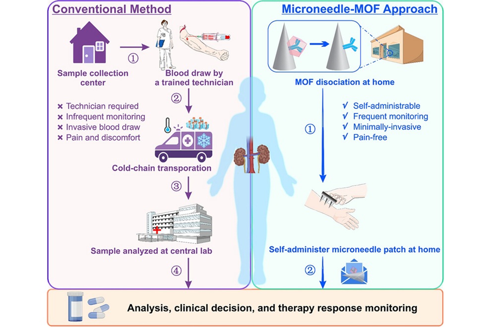
Microneedle Patch Enables At-Home Monitoring of Acute Kidney Injury
Kidney disease is often silent in its early stages, making timely detection difficult outside clinical settings. Early biomarkers such as neutrophil gelatinase–associated lipocalin can require blood draws... Read more
Business
view channel
Steris to Expand Surgical Safety Portfolio With $2.1 Billion Xodus Medical Deal
Steris has signed a definitive agreement to acquire Xodus Medical, a specialty surgical products business based in Pennsylvania, for $2.1 billion at closing. The acquisition will expand Steris’s healthcare... Read more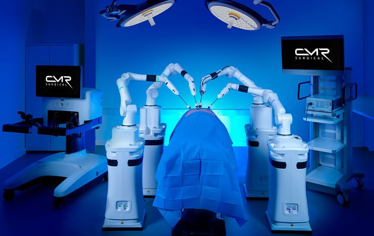
Stryker Enters Soft-Tissue Robotics Through Strategic Alliance With CMR Surgical
Stryker (Portage, MI, USA) and CMR Surgical (Cambridge, UK) have announced a strategic alliance centered on the Versius Surgical Robotic portfolio. The agreement includes an approximately USD 400 million... Read more
Terumo Neuro Acquisition Advances Liquid Embolic Technology for Chronic Subdural Hematoma
Terumo Neuro (Aliso Viejo, CA, USA) signed an agreement to acquire Arsenal Medical Inc. (Waltham, MA, USA), a privately held, clinical-stage biomaterials company. The proposed acquisition would add NeoCast,... Read more