New 3D-Printable Materials for Reconstructive Surgery Can Be Monitored Using X-Ray or CT 
|
By HospiMedica International staff writers Posted on 23 Feb 2024 |
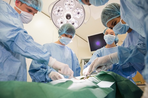
Over the past decade, gelatin-based materials have captured significant attention in research due to their simplicity in production, non-toxic nature, affordability, biodegradability, and crucially, their ability to facilitate cell growth. These attributes make them highly suitable for use in plastic and reconstructive surgery. When a surgeon implants such material into a wound, the body progressively decomposes it and substitutes it with its own tissue. This process not only expedites wound healing but also allows for the reshaping of tissues, such as in breast reconstruction post-mastectomy. Additionally, these materials are valuable for 3D printing custom implants for individual patients. Despite these advantages, there has been a significant challenge: tracking the degradation of these materials in the body has been problematic with traditional imaging techniques.
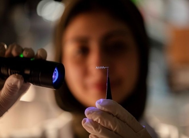
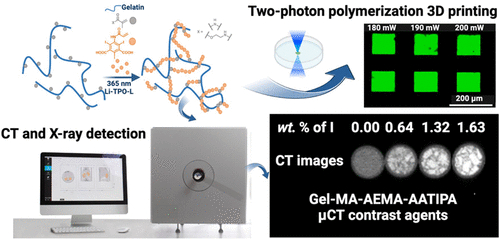
Researchers from IOCB Prague (Prague, Czech Republic) and Ghent University (Ghent, Belgium) have been refining the properties of gelatin-based materials and have introduced 3D-printable materials that are easily trackable using X-ray machines or computed tomography (CT). By incorporating a radiopaque (X-ray-contrast) agent, they have enabled the observation of the rate at which implants shrink and whether they sustain any damage. A whole series of academic papers is being written on this topic. The initial paper introduced a gelatin-based material visible via magnetic resonance imaging (MRI). The most recent publication describes materials that are detectable with X-ray and CT imaging.
This advancement allows for the ongoing monitoring of these implants, observing their biodegradation and identifying potential mechanical failures. Such data are immensely valuable in clinical settings. Leveraging this information, the biodegradation of implants can be customized to align with specific clinical needs. This is vital because tissue growth rates vary across the human body, necessitating the adaptation of implant properties accordingly. The ultimate aim is to align the biodegradation rate of these implants with the growth rate of healthy tissue. In light of these innovations, the two collaborating institutions have filed a joint patent application for the use of these materials in plastic and reconstructive surgery.
Related Links:
IOCB Prague
Ghent University
Latest General/Advanced Imaging News
Channels
Critical Care
view channel
CPR Guidelines Updated for Pediatric and Neonatal Emergency Care and Resuscitation
Cardiac arrest in infants and children remains a leading cause of pediatric emergencies, with more than 7,000 out-of-hospital and 20,000 in-hospital cardiac arrests occurring annually in the United States.... Read more
Ingestible Capsule Monitors Intestinal Inflammation
Acute mesenteric ischemia—a life-threatening condition caused by blocked blood flow to the intestines—remains difficult to diagnose early because its symptoms often mimic common digestive problems.... Read more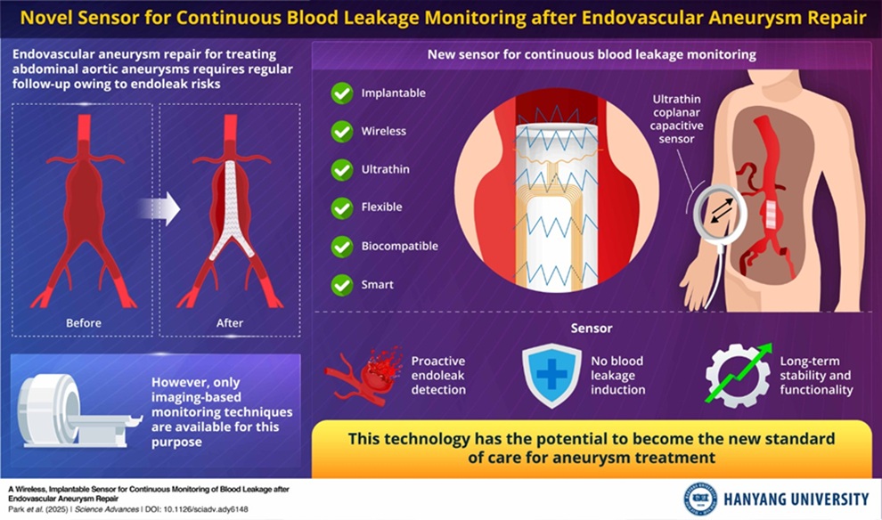
Wireless Implantable Sensor Enables Continuous Endoleak Monitoring
Endovascular aneurysm repair (EVAR) is a life-saving, minimally invasive treatment for abdominal aortic aneurysms—balloon-like bulges in the aorta that can rupture with fatal consequences.... Read more
Wearable Patch for Early Skin Cancer Detection to Reduce Unnecessary Biopsies
Skin cancer remains one of the most dangerous and common cancers worldwide, with early detection crucial for improving survival rates. Traditional diagnostic methods—visual inspections, imaging, and biopsies—can... Read moreSurgical Techniques
view channel
Robotic Assistant Delivers Ultra-Precision Injections with Rapid Setup Times
Age-related macular degeneration (AMD) is a leading cause of blindness worldwide, affecting nearly 200 million people, a figure expected to rise to 280 million by 2040. Current treatment involves doctors... Read more
Minimally Invasive Endoscopic Surgery Improves Severe Stroke Outcomes
Intracerebral hemorrhage, a type of stroke caused by bleeding deep within the brain, remains one of the most challenging neurological emergencies to treat. Accounting for about 15% of all strokes, it carries... Read morePatient Care
view channel
Revolutionary Automatic IV-Line Flushing Device to Enhance Infusion Care
More than 80% of in-hospital patients receive intravenous (IV) therapy. Every dose of IV medicine delivered in a small volume (<250 mL) infusion bag should be followed by subsequent flushing to ensure... Read more
VR Training Tool Combats Contamination of Portable Medical Equipment
Healthcare-associated infections (HAIs) impact one in every 31 patients, cause nearly 100,000 deaths each year, and cost USD 28.4 billion in direct medical expenses. Notably, up to 75% of these infections... Read more
Portable Biosensor Platform to Reduce Hospital-Acquired Infections
Approximately 4 million patients in the European Union acquire healthcare-associated infections (HAIs) or nosocomial infections each year, with around 37,000 deaths directly resulting from these infections,... Read moreFirst-Of-Its-Kind Portable Germicidal Light Technology Disinfects High-Touch Clinical Surfaces in Seconds
Reducing healthcare-acquired infections (HAIs) remains a pressing issue within global healthcare systems. In the United States alone, 1.7 million patients contract HAIs annually, leading to approximately... Read moreHealth IT
view channel
Printable Molecule-Selective Nanoparticles Enable Mass Production of Wearable Biosensors
The future of medicine is likely to focus on the personalization of healthcare—understanding exactly what an individual requires and delivering the appropriate combination of nutrients, metabolites, and... Read moreBusiness
view channel
Philips and Masimo Partner to Advance Patient Monitoring Measurement Technologies
Royal Philips (Amsterdam, Netherlands) and Masimo (Irvine, California, USA) have renewed their multi-year strategic collaboration, combining Philips’ expertise in patient monitoring with Masimo’s noninvasive... Read more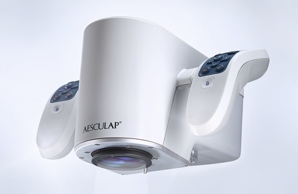
B. Braun Acquires Digital Microsurgery Company True Digital Surgery
The high-end microsurgery market in neurosurgery, spine, and ENT is undergoing a significant transformation. Traditional analog microscopes are giving way to digital exoscopes, which provide improved visualization,... Read more
CMEF 2025 to Promote Holistic and High-Quality Development of Medical and Health Industry
The 92nd China International Medical Equipment Fair (CMEF 2025) Autumn Exhibition is scheduled to be held from September 26 to 29 at the China Import and Export Fair Complex (Canton Fair Complex) in Guangzhou.... Read more