


New Microscopy Technique Enables Rapid Tumor Analysis by Surgeons in OR
|
By HospiMedica International staff writers Posted on 19 Dec 2024 |


|
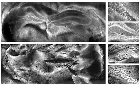
The current standard method for quickly sampling and imaging tissue during surgery involves taking a biopsy, freezing the sample, staining it to enhance visibility, and slicing it into thin sections that are then mounted on glass slides. An optical microscope is then used to examine the tissue in detail. If tumor cells are found on the surface of the tissue sample, it indicates that the surgeon has cut through, rather than around, the tumor, leaving some of the tumor behind, which may require follow-up surgery to remove more tissue. However, frozen section pathology faces issues such as tissue artifacts and reduced staining quality, which can affect diagnostic accuracy and surgical decisions. A new imaging technology now offers a faster and more cost-effective way for surgeons to image tissue samples during surgery to determine whether the entire tumor has been removed or if more tissue needs to be excised.
Bioengineers at the California Institute of Technology (Caltech, Pasadena, CA, USA) have introduced a new imaging technique called parallel ultraviolet photoacoustic microscopy (PUV–PAM), detailed in a study published in Science Advances. This method is based on photoacoustic microscopy (PAM), a technique that excites tissue samples with a low-energy laser, causing the tissue to vibrate. The system detects the ultrasonic waves emitted by the vibrating tissue. Since cell nuclei absorb more light than the surrounding material, PAM can reveal the size and distribution of nuclei and the packing density of cells. Cancerous tissue typically has larger nuclei and more densely packed cells. The research team has previously developed PAM systems for imaging bone and breast tissue, but to make these systems suitable for use in the operating room, they needed to overcome the limitations of the ultraviolet lasers used, which previously restricted the imaging speed.
To address this issue, the researchers divided a single laser beam into eight smaller, parallel laser "spots," allowing the system to cover the tissue sample more quickly. Additionally, PUV-PAM combines two scanning techniques to achieve faster imaging of slide-free tissues. These innovations make the technique approximately 40 times faster than the previous state-of-the-art methods developed by the team. The new PUV–PAM technique can eliminate the need to freeze, section, or stain tissue samples. Even thick samples with irregular surfaces, which are typically too thick for traditional microscopy, can be directly imaged. This method could enable oncologists to analyze biopsy samples during surgery, allowing them to remove additional tissue if necessary, potentially eliminating the need for follow-up surgeries.
"We hope this new imaging system can provide more opportunities for intraoperative pathological examination of slide-free specimens in oncology surgeries. We believe it has the potential to revolutionize intraoperative histology," says Rui Cao, lead author of the new paper. "With the current system, we can image a 1 cm2 sample at 1.3 micron resolution within about five minutes. And we demonstrate in the paper that this technique is effective in a variety of tissue types."





Latest Pathology News
Channels
Artificial Intelligence
view channel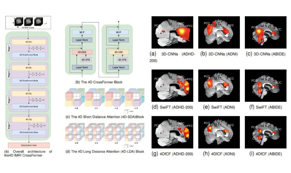
Explainable AI Tool Improves Brain Disorder Screening from 4D fMRI
Functional magnetic resonance imaging (fMRI) captures changes in brain activity over time, but the resulting image sequences can be difficult to analyze. Screening for brain disorders requires assessing... Read more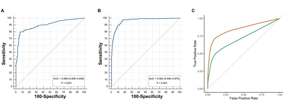
AI Improves Non-Contrast CT Interpretation for Time-Sensitive Stroke Assessment
Acute ischemic stroke occurs when a blood vessel in the brain becomes blocked, requiring rapid diagnosis to enable timely reperfusion therapy. Emergency departments often use computed tomography angiography... Read more
Critical Care
view channel
New Drug Delivery System Extends Release of Local Anesthetic for Weeks
Local anesthetics used for nerve blocks often provide pain relief for only a limited period, while perioperative pain can persist much longer. Extending the duration of a nerve block therefore requires... Read more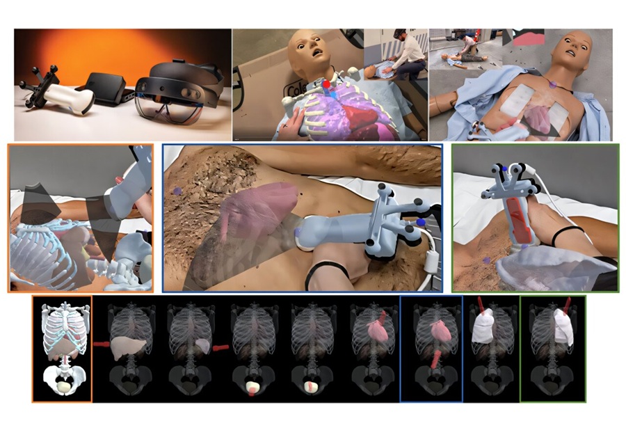
Anatomical Guidance System Improves Trauma Ultrasound Acquisition by Novice Users
Focused assessment with sonography for trauma (FAST) helps clinicians detect free fluid and signs of internal bleeding in injured patients, but obtaining reliable images can be difficult for inexperienced operators.... Read more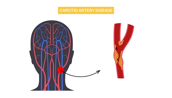
AI-Enhanced Handheld Ultrasound Improves Carotid Plaque Detection
Handheld ultrasound can miss small or faint carotid plaque, creating uncertainty in community screening. This gap makes it harder for frontline clinicians to decide who needs confirmatory imaging, closer... Read more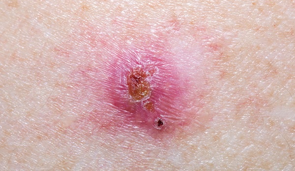
Noninvasive Imaging Approach Aims to Detect Basal Cell Carcinoma Before It Becomes Visible
Basal cell carcinoma is the most common form of skin cancer and can damage nearby structures such as the nose or eyes when it develops on the face. Diagnosis typically depends on visible skin changes,... Read moreSurgical Techniques
view channel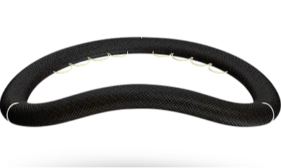
First Human Implant Performed With New Open Mitral Annuloplasty Ring
In mitral valve repair, clinicians aim to preserve the mitral annulus’s three-dimensional motion while providing enough support for effective remodeling. Ring preferences vary between open and closed ... Read more
Handheld Device Improves Targeted Cell Collection for Early Ovarian Cancer Assessment
High-grade serous ovarian cancer is the most common form of ovarian cancer, and many cases are thought to originate in the fallopian tubes. Because precursor lesions can be microscopic, they are difficult... Read more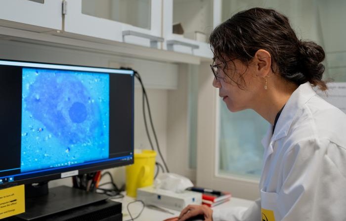
Light-Activated Nanoparticles May Offer New Approach to Retinal Prostheses
Retinitis pigmentosa is a degenerative retinal disorder in which photoreceptors progressively die, reducing visual signals to the brain while leaving surviving inner retinal circuits underused.... Read more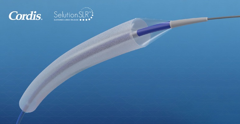
FDA Approves First Sirolimus Drug-Eluting Balloon for Coronary In-Stent Restenosis
Coronary in-stent restenosis (ISR) accounts for about 10% of the nearly one million percutaneous coronary interventions performed annually in the United States. Treating recurrent narrowing without another... Read morePatient Care
view channel
EHR Model Flags High-Risk Periods in Patients with Metastatic Breast Cancer
Determining when patients with metastatic breast cancer are nearing the end of life remains difficult, often leading to intensive interventions with limited benefit. Prognostic uncertainty can delay goals-of-care... Read more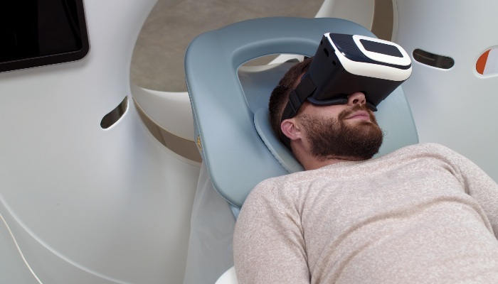
Virtual Reality Helps Claustrophobic Patients Complete Cardiac PET/CT Scans
Anxiety and claustrophobia—intense fear of confined spaces—often prevent patients from completing cardiac positron emission tomography/computed tomography (PET/CT). Aborted scans delay diagnosis, consume... Read more

Medical Imaging
view channel
New MRI Approach Captures Rapidly Decaying Tissue Signals
MRI measures signals generated as excited proton spins return toward equilibrium. However, some tissue signals decay so rapidly that conventional systems can miss them during the brief interval after excitation,... Read more
AI Tool Integrates MRI and Ultrasound to Detect Endometriosis
Endometriosis is a chronic condition in which tissue similar to the uterine lining grows outside the uterus, causing pain, heavy bleeding, and infertility. Diagnosis often depends on surgical visualization,... Read more

Health IT
view channel
New Tablet Platform Extends Stroke Neurorehabilitation Beyond the Clinic
Continuity and intensity of neurorehabilitation after stroke are critical for functional recovery, but access to ongoing therapy at home often remains limited. Hospitals and outpatient programs therefore... Read more
Smartphone App Improves Blood Pressure Control in Hypertension
Hypertension, or high blood pressure, is a major modifiable driver of cardiovascular, renal, and cerebrovascular morbidity. Despite effective therapies, many adults fail to achieve control, leading to... Read more

Point of Care
view channel
Continuous Glucose Monitoring Identifies Cardiometabolic Risk in Adults Without Diabetes
Dysglycemia—abnormal blood glucose regulation—can fluctuate throughout the day and often escape conventional screening. Clinicians typically rely on fasting plasma glucose and hemoglobin A1c, which offer... Read more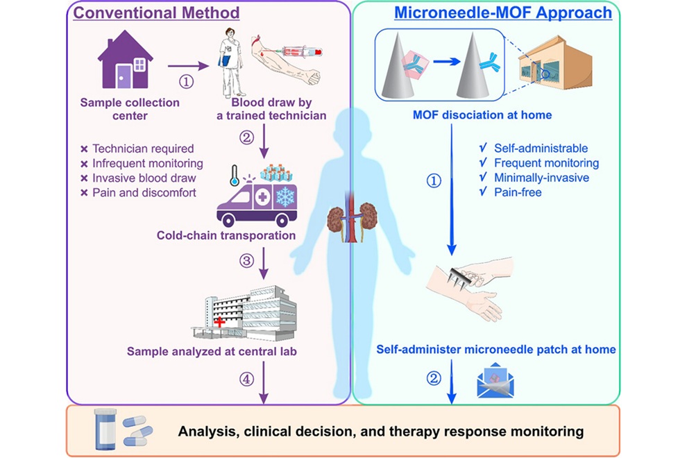
Microneedle Patch Enables At-Home Monitoring of Acute Kidney Injury
Kidney disease is often silent in its early stages, making timely detection difficult outside clinical settings. Early biomarkers such as neutrophil gelatinase–associated lipocalin can require blood draws... Read more
Business
view channel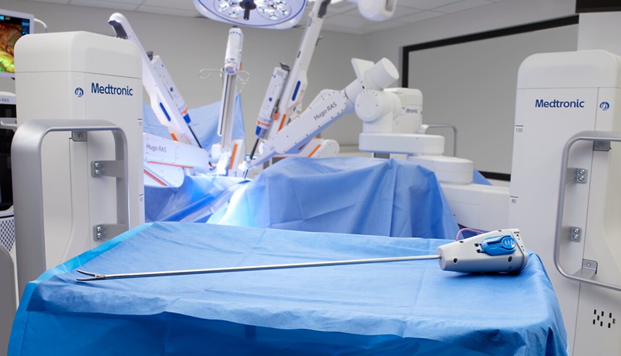
Medtronic Receives FDA Clearance for Vessel-Sealing Instrument for Robotic Surgery
As robotic-assisted surgery expands across U.S. hospitals, teams increasingly seek energy instruments with the familiarity and performance of tools used in open and laparoscopic procedures.... Read more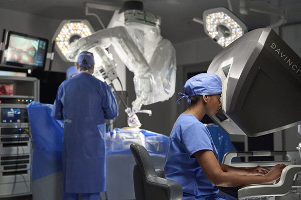
Robotic Single-Port System Gains CE Mark for Transvaginal Gynecologic Procedures
Intuitive (Sunnyvale, CA, USA) announced that it has received CE mark approval for use of the da Vinci SP Single Port surgical system in transvaginal gynecologic procedures, marking the first such indication... Read more