X-Ray Imaging System Enables Scientists to See in Real Time How Effective Treatments are for Cystic Fibrosis
By HospiMedica International staff writers
Posted on 25 Aug 2014
A new imaging approach allows researchers to monitor the effectiveness of a treatment for the life-threatening genetic disorder. Posted on 25 Aug 2014
Cystic fibrosis affects many of the body’s systems, but most severely the lungs, and currently it can take several months to measure how effective treatment is for the early-fatal lung disease.
.")
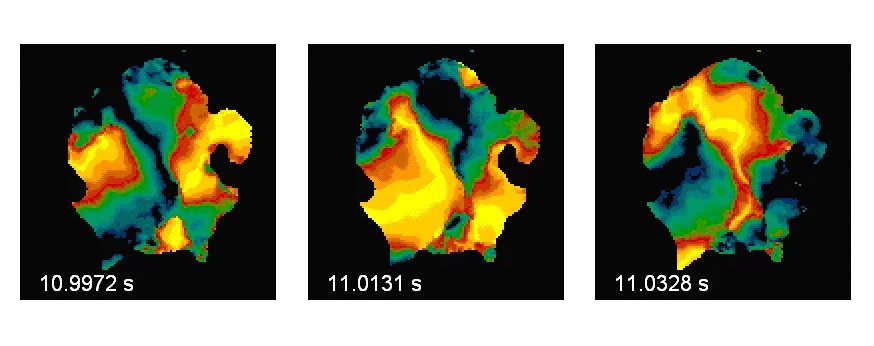
Image: Dr. Kaye Morgan, Monash University (Photo courtesy of Monash University).
Dr. Kaye Morgan, from Monash University (Melbourne; VIC, Australia), and lead researcher of the study, reported that the new X-ray imaging strategy allows researchers to visualize soft tissue structures, for example, the airways, brain, and lungs, which are effectively hidden in standard X-ray images. “At the moment we typically need to wait for a cystic fibrosis treatment to have an effect on lung health, measured by either a lung CT [computed tomography] scan or breath measurement, to see how effective that treatment is,” Dr. Morgan said. “However the new imaging technique allows us for the first time to noninvasively see how the treatment is working ‘live’ on the airway surface.”
Dr. Morgan noted that this X-ray imaging method would enable clinicians and researchers to measure how effective treatments are, and progress new treatments to the clinic at a much quicker rate, a key goal of the investigators. “Because we will be able to see how effectively treatments are working straight away, we’ll be able to develop new treatments a lot more quickly, and help better treat people with cystic fibrosis,” Dr. Morgan said.
Dr. Morgan noted that the new imaging technology, which was developed using a synchrotron X-ray source, may also create new avenues for assessing how effective treatments were for other lung, heart, and brain diseases.
The study’s findings were published August 15, 2014, in the American Journal of Respiratory and Critical Care Medicine.
Related Links:
Monash University

Gold Member
Real-Time Diagnostics Onscreen Viewer
GEMweb Live

Gold Member
12-Channel ECG
CM1200B

Silver Member
Compact 14-Day Uninterrupted Holter ECG
NR-314P

New
Ultrasound System
Voluson Signature 18