Novel Electrode Set Makes Brain Function Measurement Easier
By HospiMedica International staff writers
Posted on 09 Oct 2014
A novel electroencephalography (EEG) electrode set that is placed below the hairline delivers reliable results without any special skin preparation.Posted on 09 Oct 2014
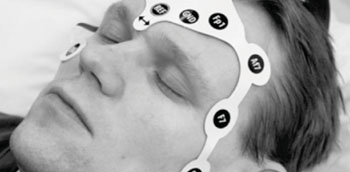
Developed at the University of Eastern Finland (Joensuu, Finland), the EEG electrode set consists of 16 hydrogel-coated electrodes which, unlike the traditional method, are placed on the hair-free areas of the patient's head. This significantly speeds up the measurement process, since there is no need to scrape the patient's skin or to use gels for adhesion. And as the electrode set is flexible and solid, the electrodes are automatically oriented to their correct locations. Furthermore, there is no need to move the patient's head when placing the EEG set, especially important in those possibly suffering from a neck or skull injury.

Image: Printed EEG electrode set measures electrical activity of the brain (Photo courtesy Pasi Lepola).
The EEG electrode set is manufactured using screen printing technology, with the conductors and measurement electrodes printed with silver ink on a flexible polyester film. Thanks to the materials used, the electrode set does not interfere with any magnetic resonance imaging (MRI) or computed tomography (CT) procedures the patient may need. The set is also disposable, and so particularly well-suited to be used in emergency care, ambulances, or even in field conditions. The performance of the electrode set was tested on volunteers and real patient cases, with the results comparable to those obtained by traditional EEG methods.
“The EEG recordings revealed that in spite of skin-electrode impedances being higher, the signal quality was comparable with that obtained with traditional cup electrodes and the clinical question could be answered accurately in almost all patient cases,” wrote Pasi Lepola, MSc, who developed the EEG electrode set for his PhD study. “The sophisticated screen-printed electrode structure with adhesive hydrogels enables stepwise attachment of electrode set within a few minutes.”
EEG is most often used to diagnose epilepsy, which causes obvious abnormalities. It can also be used to diagnose sleep disorders, coma, encephalopathy, and brain death. Despite limited resolution, EEG is a valuable tool for research and diagnosis, especially when millisecond-range temporal resolution (not possible with CT or MRI) is required.
Related Links:
University of Eastern Finland


