Injectable Agent Makes Cancerous Cells Visible During Surgery
By HospiMedica International staff writers
Posted on 01 Feb 2016
Results from a trial published on January 6, 2015, in the journal Science Translational Medicine show how a new injectable agent can be used during soft-tissue sarcoma or breast cancer surgery to identified remnants of cancerous tissue.Posted on 01 Feb 2016
Cancer surgeons use Magnetic Resonance Imaging (MRI) and Computed Tomography (CT) scans as guides during excision of tumors and surrounding tissue, however traces of cancerous tissue around the tumor remain undetected and as a result are not removed, leading to additional surgery later, and radiation therapy.
.")
Image: The injectable agent, a blue liquid called LUM015, can be used to identify cancerous tissue in human patients without adverse effects. (Photo courtesy of Shawn Rocco/ Duke Medicine).
The trial was carried out at Duke University Medical Center (Durham NC, USA) and included 15 patients undergoing surgery for soft-tissue sarcoma or breast cancer. The researchers tested the new injectable agent called LUM015 that makes tumor cells visible by causing fluorescence, and enables surgeons to find and take out all cancerous tissue in one operation.
The research into the new imaging technology was made possible by a collaboration between scientists at Duke University Medical Center, Massachusetts Institute of Technology (MIT; Cambridge, MA, USA), and a company called Lumicell (Wellesley, MA, USA).
As a next step, researchers at Massachusetts General Hospital are evaluating the efficacy and safety of LUM015 and the imaging technology in a trial with 50 women suffering from breast cancer. After that, the researchers plan to verify that the technology can reduce the number of second operations following surgery for the removal of breast cancer.
David Kirsch, MD, PhD, professor at Duke University School of Medicine, and senior author of the study, said. “At the time of surgery, a pathologist can examine the tissue for cancer cells at the edge of the tumor using a microscope, but because of the size of cancer it’s impossible to review the entire surface during surgery. The goal is to give surgeons a practical and quick technology that allows them to scan the tumor bed during surgery to look for any residual fluorescence.”
Related Links:
Duke University Medical Center
Massachusetts General Hospital
Lumicell

Gold Member
Disposable Protective Suit For Medical Use
Disposable Protective Suit For Medical Use

Gold Member
Real-Time Diagnostics Onscreen Viewer
GEMweb Live

Silver Member
Compact 14-Day Uninterrupted Holter ECG
NR-314P
New
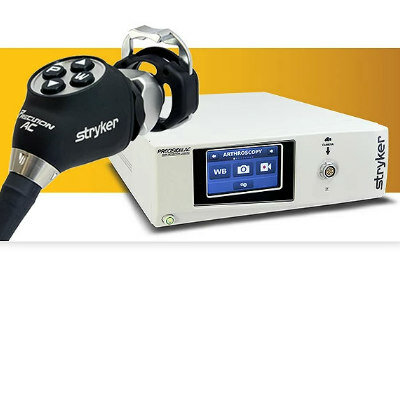
Autoclavable Camera System
Precision AC