Microneedle Skin Patch Collects Fluid for Diagnostic Testing
By HospiMedica International staff writers
Posted on 18 Jun 2019
A new study describes how a paper-based skin patch that contains tiny needles painlessly collects interstitial fluid (ISF) for biomarker analysis.Posted on 18 Jun 2019
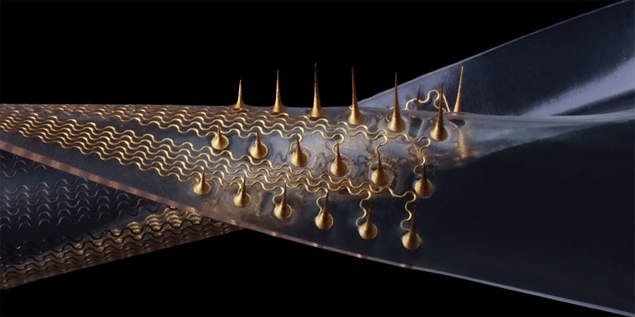
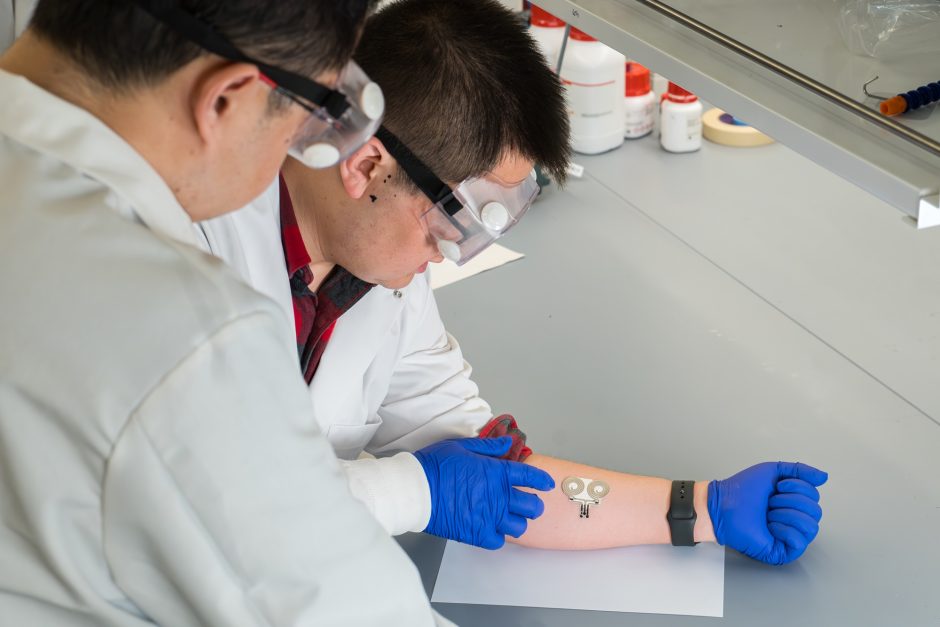
Developed at Washington University (WUSTL; St. Louis, MO, USA) and the Georgia Institute of Technology (Georgia Tech; Atlanta, GA, USA), the patch’s micrometer-scale microneedles puncture the skin, creating tiny micropores in the surface through which small quantities of ISF leak. The ISF is subsequently collected onto a strip of plasmonic paper that forms part of the backing of the patch. It is then analyzed in-situ, using surface-enhanced Raman scattering (SERS), in order to detect and measure pharmacokinetic profiles.
.")
Tiny microneedles help analyze ISF biomarkers (Photo courtesy of ACS).
For the study, the researchers immobilized negatively charged poly(styrenesulfonate) (PSS) coated gold nanorods on a thin strip of filter paper using plasmonic calligraphy. A positively charged dye containing rhodamine 6G (R6G) was then injected into a rats' bloodstream. The dye entered the ISF and from there to the microneedle patch and onto the plasmonic paper, where it bound to the negatively charged PSS, and then analyzed with SERS. The researchers found that the new method could detect the R6G dye as sensitively as previous multi-step procedures. The study was published on May 9, 2019, in ACS Sensors.
“This proof-of-concept study indicates that a plasmonic paper microneedle patch has the potential to enable on-patch measurement of molecules in ISF for research and future medical applications,” concluded lead author Chandana Kolluru, PhD, of the Georgia Tech School of Materials Science and Engineering, and colleagues.
ISF surrounds all tissue cells and is present in the skin. It is advantageous for biosensing applications since it does not contain any particulates (red blood cells or platelets), and contains at least 5–10 times less protein than blood serum. However, only extremely low volumes can be found on the skin, making the process of ISF extraction rather difficult. ISF extraction using microneedles combined with integrated biosensing capabilities could provide significant opportunities for minimally invasive monitoring and diagnostics, such as in diabetes.
Related Links:
Washington University
Georgia Institute of Technology

Gold Member
POC Blood Gas Analyzer
Stat Profile Prime Plus

Gold Member
SARS‑CoV‑2/Flu A/Flu B/RSV Sample-To-Answer Test
SARS‑CoV‑2/Flu A/Flu B/RSV Cartridge (CE-IVD)

Silver Member
Wireless Mobile ECG Recorder
NR-1207-3/NR-1207-E
New
Electric Bariatric Patient Lifter
SVBL 205