Imaging Technology Illuminates Lung Cancer Tumors to Make Surgical Removal Easier
Posted on 28 Jun 2022
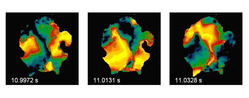
Non-small-cell lung cancers (NSCLC), often related to smoking, are the most common form of lung cancer and can be life threatening. Now, a study aims to further advance an intraoperative imaging technology that illuminates tumor tissue and can make it easier and safer for both clinicians and patients to remove tumors on or in the lungs. The technology, intraoperative molecular imaging (IMI), is based on fluorescent beacon molecules that target and bind themselves to tumor cells, essentially making them glow - and allowing doctors to more easily distinguish cancer from healthy tissue.
Researchers at Penn Medicine (Philadelphia, PA, USA) have received a five-year, USD 9 million research grant from the National Cancer Institute (NCI) to study and improve the IMI technology for NSCLC. The new grant-funded research project aims to develop improved beacon molecules for NSCLC and imaging equipment to go with it, then test them in clinical trials.
")
The fluorescent beacon molecules used in IMI are normally infused into the patient hours or days before surgery. They bind to cell-surface receptors, such as folate receptors, which are particularly abundant on cancer cells. The light the beacons emit is typically in the near-infared range, allowing for visualization detection of tumor cells up to about two centimeters below the tissue surface, depending on the tissue type. Tissue tagged with these fluorescing beacons can be imaged in real-time, during surgery, with relatively inexpensive and portable equipment.
Data from additional clinical trials have shown it also has the potential to help doctors detect tumors - for example, following a positive or ambiguous X-ray finding - during non-surgical inspections of patients’ lungs via bronchoscopy, when doctors use a scope to investigate the passages in a person’s lungs.
“This funding gives us a tremendous opportunity to further evaluate this important technology and with the goal being to improve outcomes for patients,” said grant principal investigator Sunil Singhal MD, the William Maul Measey Professor in Surgical Research and Chief of Thoracic Surgery, and director of the Center for Precision Surgery in the Abramson Cancer Center at Penn. “We aim to develop this technology even further and to study it in additional clinical trials to help improve surgical identification and removal of tumors.”
“Complete resection is the best outcome for patients, and the goal in this program is to improve the chances of achieving that without unnecessary tissue removal,” said Ronald DeMatteo MD, the John Rhea Barton Professor of Surgery and Chair of the Department of Surgery at Penn.
Related Links:
Penn Medicine