Novel Method Combining Heart Biopsy and Device Implantation Reduces Complications Risk
Posted on 22 Feb 2025
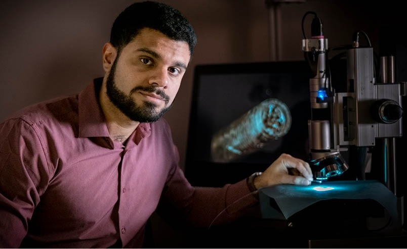
Endomyocardial biopsy (EMB) is a crucial diagnostic tool for identifying various cardiac conditions; however, it carries a risk of complications due to its invasive nature. New research has introduced a method that combines right ventricular (RV) septal EMB with the implantation of cardiac implantable electronic devices (CIEDs) such as pacemakers or defibrillators, utilizing the same sheath for both procedures. This innovative approach, detailed in two studies published in the journal Heart Rhythm, highlights the feasibility and safety of the technique.
EMB is associated with a complication risk of up to 3%. It is typically performed only when noninvasive diagnostic methods fail to provide definitive results or the necessary histology, which can result in delays. The newly developed combined procedure significantly reduces the risk of complications and allows for earlier diagnosis of heart conditions such as cardiomyopathy and heart failure. Early detection enables timely treatment, which can improve patient outcomes. Furthermore, it can help identify genetic factors that may affect other family members. The studies published in Heart Rhythm discuss the researchers’ experience in combining RV septal EMB with CIED implantation, taking advantage of 3D curved conduction system pacing (CSP) sheaths. These CSP sheaths allowed for precise RV septal placement, making the biopsy procedure a quick and safe addition to the CIED implantation process.
")
"Our study ‘Novel technique and assessment of available 3-dimensional delivery sheath for endomyocardial biopsy during cardiac device implantation’ demonstrated the usefulness and safety of the new method for performing EMB in 20 patients using a 3-dimensional delivery sheath during CIED implantation,” said Kenji Kuroki, MD, Department of Cardiovascular Medicine, University of Yamanashi (Chuo, Japan). “Additionally, it enabled the early diagnosis of previously undetected cardiac amyloidosis (CA) in 4 patients (20%), as confirmed by pathologic findings. Subclinical CA may often be overlooked among patients with CIED implantation. Early diagnosis could enable the timely initiation of appropriate treatment, making this finding particularly important."
Patients requiring CIEDs often experience complications such as cardiomyopathy or other cardiac issues, which may necessitate EMB. Traditionally, EMB using a bioptome—a tool for extracting tissue from the heart—has been avoided in these patients to minimize the risk of lead displacement during the peri-procedural period, in accordance with EMB guidelines. The study titled "Feasibility and safety of endomyocardial biopsy by lumenless pacing lead sheath–directed method during cardiac implantable electronic device implantation" enrolled 80 patients who were referred for EMB. This study evaluated the safety and practicality of performing transvenous RV EMB using the lead sheath method (L-S-M) during CIED implantation and compared it to the traditional bioptome method (T-B-M).
"Using cardiac pacing leads instead of traditional myocardial biopsy forceps during the implantation could be timesaving and cost-effective without compromising safety,” said Yang Ye, lead investigator at Zhejiang University (Hangzhou, China). “In addition, it has the advantages of requiring only one vessel for both EMB and CIED implantation. In this study, no ventricular perforations or serious cardiac event occurred, demonstrating a safe L-S-M procedure of EMB."