Optical Device Offers Less Painful Colonoscopy
By HospiMedica International staff writers
Posted on 09 May 2011
A colonoscopy device under development could help reduce patient discomfort while still ensuring the accuracy of the exam procedure.Posted on 09 May 2011
Researchers at Tufts University (Medford, MA, USA) developed endoscopic fiber optic shape tracker (EFOST) technology as a possible solution to the problem that occurs when the endoscope is inserted into the colon during a routine screening. As the endoscope is navigated through the bends and turns in the colon, its tip can impinge against the colon wall. When this happens, the tip turns stationary; if the physician applies more pressure, a loop can form in the length of scope behind the tip. Since the traditional endoscope provides only a frontal view during the procedure, the doctor cannot see the loop, nor easily maneuver the scope to remove it. As a result, looping can be a major source of pain during a colonoscopy.

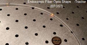
Image: Quantum dots lighting up with the EFOST (Photo courtesy of Tufts University).
The researchers developed a prototype to overcome this problem, by embedding quantum dots--nano-sized crystals of semiconductor material--circumferentially at intervals along the length of an optical fiber. They then stretched the fiber around a metal cylinder to create a bending effect, and injected a laser light beam into the fiber's inner core from one end; the fiber's core released light as it is bent, activating the quantum dots. Instantly, the dots reemitted light signals of varying intensity to a spectrometer; using this data, the researchers were able to measure the degree of curvature in the fiber. And from the position of the activated dots, the researchers were also able to calculate the direction of the bend.
"Doctors will have a way to see in real-time how the scope is moving inside the body. If the scope begins to loop, they will see it instantaneously and then be able to make adjustments to straighten it out,” said lead researcher associate professor of mechanical engineering Caroline Cao, PhD. "Physicians can use the image on the monitor to guide them. They'll know exactly where the end of the point is, as well as the shape of the scope inside the colon.”
Colonoscopy is the examination of the colon and the distal part of the small bowel with a video camera or a fiber optic camera on a flexible endoscope passed through the anus. It may provide a visual diagnosis (e.g., ulceration, polyps) and grants the opportunity for biopsy or removal of suspected lesions. Virtual colonoscopy, which uses imagery reconstructed from computed tomography (CT) scans or from nuclear magnetic resonance (MR) scans, is also possible, as a totally noninvasive medical test, although it is not standard and still under investigation regarding its diagnostic abilities.
Related Links:
Tufts University


