New Guidance System Improves Minimally Invasive Surgery
By HospiMedica International staff writers
Posted on 28 Apr 2014
An image-based computerized guidance system could make minimally invasive surgery (MIS) more accurate and streamlined using equipment already common in the operating room (OR).Posted on 28 Apr 2014
Researchers at Johns Hopkins University (JHU; Baltimore, MD, USA) have developed an algorithm for intensity-based analysis of computerized tomography (CT) and X-ray projections to allows direct registration of preoperative CT and planning data for intraoperative fluoroscopy, thus providing three dimensional (3D) localization that is free from conventional limitations associated with external fiducial markers, stereotactic frames, trackers, and manual registration. Initial testing shows that the algorithm is potentially superior to conventional tracking systems that have been the mainstay of surgical navigation over the last decade.
.")
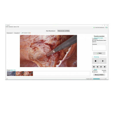
Image: JHU graduate student Ali Uneri demonstrates the new surgical guidance system (Photo courtesy of JHU - Johns Hopkins University).
From just a single X-ray frame, the researchers achieved better than 3 millimeters of accuracy. The researchers then investigated navigation using two close images; they found that as little as 15 degrees of angulation was sufficient to provide 2 millimeters of accuracy, consistently better than a conventional surgical tracker. An added advantage of the system is that the two X-ray images can be acquired at an extremely low dose of radiation, far below what is needed for a visually clear image, but enough for the algorithm to extract accurate geometric information. The study was published in the December 2013 issue of Physics in Medicine and Biology.
“Imaging in the operating room opens new possibilities for patient safety and high-precision surgical guidance,” said professor of biomedical engineering Jeffrey Siewerdsen, PhD, of the JHU School of Medicine. “In this work, we devised an imaging method that could overcome traditional barriers in precision and workflow. Rather than adding complicated tracking systems and special markers to the already busy surgical scene, we realized a method in which the imaging system is the tracker and the patient is the marker.”
“Accurate surgical navigation is essential to high-quality minimally invasive surgery,” said A. Jay Khanna, MD, an associate professor of orthopedic surgery and biomedical engineering at JHU, who evaluated the system in its first application to spine surgery. “But conventional navigation systems can present a major cost barrier and a bottleneck to workflow. This system could provide accurate navigation with simple systems that are already in the OR and with a sophisticated registration algorithm under the hood.”
Related Links:
Johns Hopkins University

Gold Member
Solid State Kv/Dose Multi-Sensor
AGMS-DM+

Gold Member
12-Channel ECG
CM1200B

Silver Member
Compact 14-Day Uninterrupted Holter ECG
NR-314P
New
Examination Data Management Software
DiVAS 2.8