Pioneering System Diagnoses Cancerous Tissue During Endoscopy
By HospiMedica International staff writers
Posted on 16 Jun 2014
A biomedical engineering team has developed a first of its kind in vivo molecular diagnostic system that makes highly objective, real-time cancer diagnosis during endoscopic examination a reality.Posted on 16 Jun 2014
A National University of Singapore (NUS) team led by Associate Professor Huang Zhiwei, Department of Biomedical Engineering, has developed what is currently the only system clinically shown to be used in human patients for diagnosing even precancerous tissue in gastrointestinal tract during endoscopic examination in real time. Unlike conventional endoscopy that relies on the physician's visual interpretation of the images followed by a pathologist's analysis of the biopsy specimen several days later, their diagnostic system utilizes computer analysis of biomolecular information that can provide diagnosis in real time. It is a paradigm shift from a complex to a simple, objective, and rapid diagnostic procedure.
.")
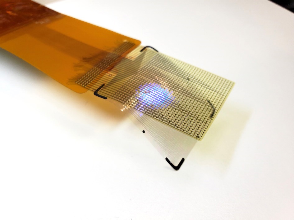
Image: Rapid fiber-optic confocal Raman spectroscopy system developed for real-time in vivo epithelial tissue diagnosis and characterization during endoscopy (Photo courtesy of Prof. Zhiwei Huang, the National University of Singapore, and the journal Gastroenterology).
The In Vivo Molecular Diagnostic (IVMD) system is based on confocal Raman spectroscopy and includes a proprietary confocal fiber-optic probe connected to a customized online software control system. The fiber-optic probe enables the collection of biomolecular fingerprint of tissues in less than a second—while the online software enables this information to be extracted and analyzed, with diagnostic result presented during endoscopic examination. The IVMD system has been used in more than 500 patients in Singapore across diverse cancer types such as stomach, esophagus, colon, rectum, head and neck, and cervix. The researchers have also published more than 40 peer-reviewed publications, most recently a report by Bergholt MS, et al. in the journal Gastroenterology, published January 2014.
“We are delighted to not only overcome the technical challenges of weak Raman signal, high fiber background noise, and lack of depth perception by using our specially designed probe, but also to enable real-time diagnostic results to be displayed during endoscopy with our customized software,” said Prof. Huang.
For the clinical testing, the team has been collaborating with researchers from the NUS Yong Loo Lin School of Medicine, led by its Dean, Associate Professor Khay Guan Yeoh. Prof. Yeoh commented, “This remarkable new system is the first such diagnostic probe that can be used real-time, inside the human body, providing almost instantaneous information on cellular changes, including cancer and pre-cancer. This is a first in the world development, pioneered here in Singapore. It has the potential to make enormous clinical impact to how cancer is diagnosed and managed. The immediate point-of-care diagnosis during live endoscopic examinations will provide benefits in time and cost-savings, and will improve our patients’ prognosis.”
“It has been a long tedious journey of more than 10 years. The journey could be longer if not for the excellent cross-disciplinary teamwork at NUS. The contribution of the NUS clinical team is invaluable in demonstrating the clinical benefits of the system,” added Prof Huang. Moving forward, the team will conduct larger scale clinical trials, mainly in gastrointestinal cancer, to further validate the utility of this novel system.
Related Links:
National University of Singapore
Video: Clinical Use of Raman spectroscopy software during an endoscopic procedure

Gold Member
POC Blood Gas Analyzer
Stat Profile Prime Plus

Gold Member
SARS‑CoV‑2/Flu A/Flu B/RSV Sample-To-Answer Test
SARS‑CoV‑2/Flu A/Flu B/RSV Cartridge (CE-IVD)

Silver Member
Wireless Mobile ECG Recorder
NR-1207-3/NR-1207-E

New
CT Phantom
CIRS Model 610 AAPM CT Performance Phantom