3D Cone Beam CT System Designed for ENT, Oral Surgeons
By HospiMedica International staff writers
Posted on 15 Sep 2014
A cone beam, three-dimensional (3D) computed tomography (CT) imaging system has been developed for modern multispecialty practices to help ear/nose/throat (ENT) and dentomaxillofacial surgeons to make accurate diagnostics, precise treatment planning for implant, ENT, and oral surgery, and perform follow-up exams. Posted on 15 Sep 2014
The Scanora 3Dx system, developed by Soredex (Tuusula, Finland), offers excellent image quality combined with low radiation doses. The design supports convenient patient positioning and easy workflow. Compared to its sister unit, the Scanora 3D, the 3Dx offers a greater variety of field-of-view (FOV) options, from small highlight scans to large, overall head and neck imaging.
.")
Image: Scanora 3Dx cone-beam CT system (Photo courtesy of Sorodex).
The technology provides higher spatial resolution, which enables the visualization of bony structures in finer detail than in conventional CT. The system has a rigid construction with small footprint.
Features of the technology include a wide variety of FOV to accommodate multiple application areas in the head and neck region in which the FOV size can be optimized to avoid radiation sensitive organs and can be freely located due to motorized movements of chair and chin rest; open software architecture allows the user to choose the best solution according to specific needs and preferences; an optional charged-coupled device (CCD)-RealPAN sensor for high quality dental panoramic imaging, with AutoSwitch 2D/3D mode change (no manual sensor change); an intuitive and simplified clear touch control panel designed for uncomplicated operation; the ability to scout images to help position the 3D volume precisely where it is needed; and the patients are seated with their heads in normal, upright position and accommodates wheel chair patients.
The system is compatible with leading image-guided surgical navigation systems (IGS), with a Digital Imaging and Communications in Medicine (DICOM)/picture archiving and communication system (PACS) compatibility for integration with third-party software and information sharing.
Related Links:
Soredex

Gold Member
Real-Time Diagnostics Onscreen Viewer
GEMweb Live

Gold Member
12-Channel ECG
CM1200B

Silver Member
Compact 14-Day Uninterrupted Holter ECG
NR-314P
New
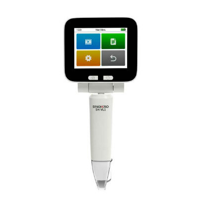
Video Laryngoscope
SH-VL1
.jpg)