Improved Ergonomics Help Neurosurgeons Stay Focused
By HospiMedica International staff writers
Posted on 30 Oct 2014
A new neurosurgical microscope enables surgeons to see better into deep, narrow cavities, allowing them to work in a neutral, upright posture which helps prevent strain and fatigue.Posted on 30 Oct 2014
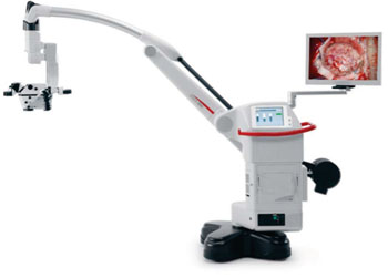
The Leica M530 OH6 is equipped with FusionOptics technology, which utilizes two separate beam paths; one path provides high resolution, the other provides depth of field. The surgeon’s brain merges the two images into one, taking the best information from the two sources. The outcome is a larger, three-dimensional (3D) area that remains in full focus, resulting in surgeons spending less time refocusing the objectives.

Image: The Leica M530 OH6 neurosurgical microscope (Photo courtesy of Leica Microsystems).
The neurosurgical microscope is provided with premium quality apochromatic optics, which together with small angle illumination (SAI) allows the beam generated by the powerful 400 Watt Xenon light to penetrate to the bottom of deep, narrow, cavities during procedures. Other features include customizable optics, binoculars with 360° rotation, an optional magnification multiplier (which increases magnification by 40%), and independent fine focusing for the rear assistant, thus increasing flexibility for surgeon and assistant alike.
Additional ergonomic benefits include a working distance of 600 millimeters and a compact optics carrier, allowing surgeons to work more comfortably and nearer to the operative site. The modular, integrated design of the Leica M530 OH6 permits surgeons to choose the options they need initially and upgrade in the future. Possible upgrades include the Leica FL400 or FL800 intraoperative fluorescence modules, as well as TrueVision 3D high-definition (HD) visualization. The Leica M530 OH6 neurosurgical microscope is a product of Leica Microsystems (Wetzlar, Germany).
“The Leica M530 OH6 takes optical quality to a new level. The instrument offers an unsurpassed view into deep cavities,” said Sandra Sokoloski, director of microsurgery at Leica Microsystems. “Through FusionOptics, surgeons also spend less time refocusing, which means less interruption to workflow. When developing this neurosurgical microscope, we wanted to enable surgeons to stay focused on their primary objective: delivering optimal results to their patients.”
“In addition to advanced optics in a more compact head, the Leica M530 OH6 offers improvements for the assistant surgeon with better positioning and fine focus controls,” said Prof. Howard A. Riina, MD, vice chairman of the department of neurosurgery and at NYU Langone Medical Center (New York, NY, USA). “Advances in image guidance integration and 3D video capabilities all maintain this microscope as the industry standard for neurosurgical operating microscopes.”
Related Links:
Leica Microsystems


