Surgical Navigation Instrument Monitors Nerve Function
By HospiMedica International staff writers
Posted on 09 Apr 2015
A surgical assistance system warns surgeons against possible injury to microstructure nerves during operations in the pelvic area. Posted on 09 Apr 2015
Researchers at the Fraunhofer Institute for Biomedical Engineering (IBMT; St. Ingbert, Germany) are developing a continuous intraoperative neuromonitoring device that involves placing flexible, wafer-thin electrodes directly onto nerve fibers and stimulating them by means of electric impulses. Tracking software then evaluates whether the surgical intervention is affecting the autonomous nerve network; if the surgeon gets too close to a nerve, or pushes or distends it, a visual and acoustic warning is emitted in case of danger of injury.
.")
Image: The continuous intraoperative neuromonitoring instrument (Photo courtesy of IBMT).
Since surgery in the pelvic area can often last several hours, the system uses dry silicone electrodes that contain nanoparticles within the silicone to guarantee the necessary conductivity; in contrast to conventional electrodes, silicone electrodes provide a stable and reliable interface over an extended period of time. The researchers are already working on a successor project, an assistance system for the stimulation of autonomous pelvic nerves and intraoperative neuromonitoring during minimally invasive surgery (MIS) and laparoscopy.
This differs from standard surgery in that electrodes have to be placed on the body externally. The problem is that the sacrum lies between the electrodes and the nerve network, obstructing the electrical field. To overcome the interference, an electrode array is placed so that it forms a grid-like field. Individual electrodes are then activated to optimize the geometry of the electric field in such a way that the surgeons can affect neuromodulation. A smart algorithm evaluates the raw signals and processes them in such a way that the surgeon can estimate risk of injury.
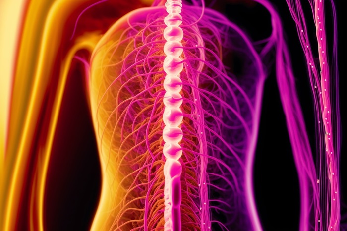
“In terms of color and structure, it’s very difficult to distinguish this nerve network from other tissue and smaller blood vessels, which frequently leads to injury,” said Prof. Klaus-Peter Hoffmann, PhD, of IBMT and the faculties of neurobiology, biology, and biotechnology at Ruhr University Bochum (Germany). “What makes it all the trickier is that often the surgeon doesn’t notice the injury during the surgery, and problems only become evident a few weeks after the operation.”
Complications of bowel surgery in the pelvic area are frequent, with more than half of the patients struggling with incontinence or sexual dysfunction due to nerve tissue damage, as the nerves controlling bladder, anus, and sexual functions all surround the intestine in a wafer-thin web.
Related Links:
Fraunhofer Institute for Biomedical Engineering
Ruhr University Bochum

Gold Member
SARS‑CoV‑2/Flu A/Flu B/RSV Sample-To-Answer Test
SARS‑CoV‑2/Flu A/Flu B/RSV Cartridge (CE-IVD)

Gold Member
Disposable Protective Suit For Medical Use
Disposable Protective Suit For Medical Use

Silver Member
Compact 14-Day Uninterrupted Holter ECG
NR-314P
New
Ultra Low Floor Level Bed
Solite Pro