3-D Printing Helps Rehearse Complex Brain Procedures
By HospiMedica International staff writers
Posted on 25 Aug 2015
A new study describes how custom, high–fidelity three-dimensional (3-D) printed models of brain vessel malformations can be used to rehearse pediatric surgical procedures. Posted on 25 Aug 2015
Researchers at Boston Children’s Hospital (BCH; MA, USA) reported on the first four cases of children benefiting from 3-D printing of their anatomy before undergoing high-risk brain procedures. The children ranged in age from two months to 16 years old; three of the four children had arteriovenous malformations (AVMs), while the fourth was a two-month-old infant with a rare vein of Galen aneurysmal malformation (VOGM), a choroidal AVM that typically results in high-output congestive heart failure or may present with developmental delay, hydrocephalus, and seizures.

3-D Printing Helps Rehearse Complex Brain Procedures
Life-sized and enlarged 3D models were created in collaboration with the BCH Hospital Simulator Program (SIMPeds), using brain magnetic resonance and arteriography data for each child, with 98% match to the children's actual anatomy. During surgery, the VOGM was embolized successfully, and all the AVMs were resected without complication. When two of the AVM patients were compared with matched controls, those with 3-D models had their surgical procedure time reduced by 30 minutes (12%). The study was published on July 31, 2015, in the Journal of Neurosurgery: Pediatrics.
“Even for a radiologist who is comfortable working with and extrapolating from images on the computer to the patient, turning over a 3-D model in your hand is transformative,” said study coauthor Darren Orbach, MD, PhD, chief of interventional and neurointerventional radiology at BCH. “Our brains work in three dimensions, and treatment planning with a printed model takes on an intuitive feel that it cannot otherwise have.”
“These children had unique anatomy with deep vessels that were very tricky to operate on. The 3-D-printed models allowed us to rehearse the cases beforehand and reduce operative risk as much as we could,” said senior author neurosurgeon Edward Smith, MD. “AVMs are high-risk cases and it's helpful to know the anatomy so we can cut the vessels in the right sequence, as quickly and efficiently as possible. You can physically hold the 3-D models, view them from different angles, practice the operation with real instruments and get tactile feedback.”
Related Links:
Boston Children’s Hospital

Gold Member
POC Blood Gas Analyzer
Stat Profile Prime Plus

New
Plate System
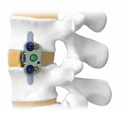
ADIRA XLIF Plate System
New
Treatment Chair
2 Section Bariatric Plinth