Tumor-Specific Imaging Agent Helps Resect Ovarian Cancer
By HospiMedica International staff writers
Posted on 29 Jun 2016
A novel contrast agent for intraoperative near-infrared (NIR) fluorescence imaging can help surgeons detect nearly 30% more ovarian tumor tissue, according to a new study.Posted on 29 Jun 2016
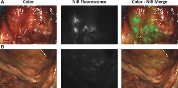
Developed by researchers at Leiden University Medical Center (LUMC, The Netherlands), the new, tumor-specific agent, called OTL38, is a combination of a NIR fluorescent dye and a folate analog. A dedicated imaging system is used to identify the fluorescent signal generated after the agent binds to a protein called folate receptor-alpha (FRα), which is expressed in more than 90% of ovarian cancers, but in much lower levels in healthy tissue. Since NIR light at 796 nm penetrates centimeters-deep into tissue, surgeons can use OTL38 to visualize tumors under the surface of the tissue.

Image: An intraoperative detection of ovarian cancer metastases using fluorescence-based imaging (Photo courtesy of LUMC).
In a randomized, double blind, placebo-controlled clinical trial, the researchers administered OTL38 to 12 patients who had epithelial ovarian cancer and were scheduled for cytoreductive surgery. They measured tolerability and blood pharmacokinetics, as well as the ability to detect the tumor. The results showed that OTL38 accumulated in both FR-α tumors and metastases, enabling the surgeons to resect an additional 29% of malignant lesions that were not identified using inspection or palpation. The study was published on June 15, 2106, in Clinical Cancer Research.
“Surgery is the most important treatment for ovarian cancer, and surgeons mainly have to rely on their naked eyes to identify tumor tissue, which is not optimal,” said lead author Alexander Vahrmeijer, MD, who heads the image-guided surgery group at LUMC. “A limitation of this study is that we cannot say yet what the impact of our findings is on cure or survival of the patients. It is reasonably plausible to assume that if more cancer is removed the survival will be better.”
Folate can be used like a Trojan horse to sneak an imaging agent or drug into a cancer cell; ovarian cancer has one of the highest rates of FR-α receptor expression. Approximately 80% of endometrial, lung, and kidney cancers, and 50% of breast and colon cancers also express the receptor.
Related Links:
Leiden University Medical Center


