Ultrasound-Based Technique to Control Myoblast Orientation Advances Tissue Engineering
Posted on 13 Dec 2024
One of the primary objectives of regenerative medicine is to develop reliable methods to replace damaged or dead tissue. With continuous progress in tissue engineering and biomedicine, we are approaching a point where growing cell sheets in the laboratory and transplanting them onto damaged organs could soon become a reality. In fact, myoblast cell sheets have already been used to successfully treat severe heart failure, showcasing the potential of this technology. Despite these advancements, there are still unresolved challenges that limit the widespread use of cell sheets in regenerative medicine. In particular, cells and their surrounding extracellular matrix must maintain specific orientations to properly perform their biological functions. This is particularly crucial in skeletal muscle fibers, which need to be aligned in a single direction. Unfortunately, when cultured using conventional methods, myoblasts—precursors to skeletal muscle cells—tend to grow in random directions. This random orientation hinders the utility of myoblast cell sheets for generating functional skeletal muscle.
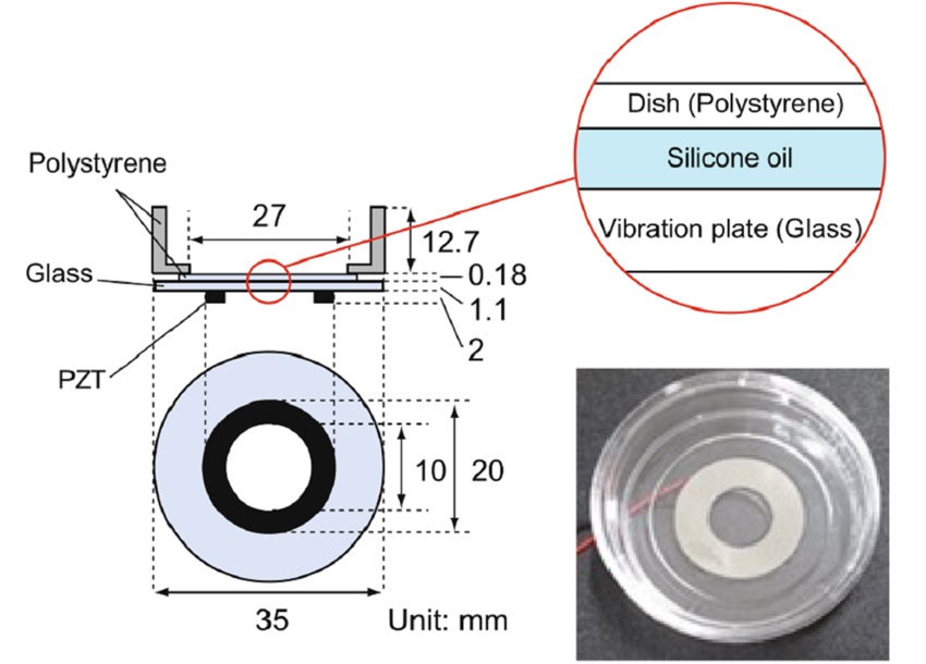
To address this, a research team from Doshisha University (Kyoto, Japan) has been exploring methods to control the orientation of cultured myoblasts. Their latest study, published in Scientific Reports, introduces an innovative technique that utilizes ultrasonication to guide cell growth in specific directions. The technique involves attaching a piezoelectric transducer to the bottom of a circular glass plate, placing a standard polystyrene cell culture dish on top, and using silicone oil as a coupling material to efficiently transmit ultrasonic vibrations to the culture surface. This simple system is placed within a chamber that regulates temperature, humidity, and CO2 concentration. The researchers use a function generator to send a sinusoidal electrical signal to the transducer. In their experiments, they applied this setup to C2C12 cells, a widely used mouse myoblast strain capable of differentiating into myotubes. The team specifically examined the effects of frequency, amplitude, timing, and duration of ultrasonication on the differentiation and orientation of cultured myoblasts.

Through careful analysis using 2D fast Fourier transform on phase-contrast images, the team quantitatively assessed the alignment of the cells and their maturation into myotubes. The results revealed an interesting correlation between cell orientation and vibrational amplitude, suggesting that the cells grew in a direction that minimized variations in displacement along their long axes. This resulted in C2C12 cells aligning circumferentially around the center of the plate. Notably, these circumferential orientations were observed even at distances greater than 4 mm, where the vibrational displacement from the ultrasonic transducer was minimal. The researchers hypothesized that this alignment could be influenced by cell-to-cell communication established between myoblasts before they fused into myotubes.
To further investigate the effects of ultrasonication, the team conducted real-time PCR and immunostaining, which revealed that the ultrasonicated cells exhibited higher levels of differentiation-related genes and characteristic morphological changes. These findings suggest that ultrasonication promotes the differentiation of myoblasts into myotubes. This technique may offer a viable alternative to or complement current chemically induced differentiation methods. Overall, the ultrasound-based approach provides a simple yet effective strategy for controlling the orientation of cultured muscle cells. However, further testing is necessary to optimize the protocol. With additional refinement, this technique could lead to innovative solutions to current challenges in regenerative medicine using cell sheets. As the technology continues to evolve, it could enable groundbreaking clinical applications in the field of cultured skeletal muscle tissues.
“The screening of different ultrasound conditions, including different frequency, output intensity, timing, and duration, will be crucial for establishing an optimized method for ultrasound-induced myotube formation in the future,” said Professor Daisuke Koyama, Faculty of Science and Engineering, Doshisha University, who led the research team.


