Neuroimaging May Help Track Brain Volume in MS Patients
By HospiMedica International staff writers
Posted on 14 Jul 2016
A new software tool could enable the accurate identification and quantification of ventricular volume in multiple sclerosis (MS), despite variability in brain shapes and scanning technology used.Posted on 14 Jul 2016
Developed by researchers at the University at Buffalo (NY, USA), the Neurological Software Tool for Reliable Atrophy Measurement in MS (NeuroSTREAM) tool estimates brain atrophy by measuring lateral ventricular volume (LVV), one of the brain structures that contain cerebrospinal fluid (CSF); when atrophy occurs, the LVV expands. Since the LVV is anatomically distinctive and strategically located in the center of the brain, focusing on it provides a metric that is relatively immune to the adverse impacts of imprecise positioning, gradient distortions, incomplete head coverage, and other motion and wraparound artifacts.
.")
Image: Brain scans of patients with the LVV highlighted in red (Photo courtesy of the University at Buffalo).
The new tool simplifies the calculation of brain atrophy based on data from routine magnetic resonance imaging (MRI) scans, by comparing them to a database bank of 20,000 brain scans taken from of MS patients. The software runs on a user-friendly, cloud-based platform that is easily available from workstations, laptops, tablets, iPads, and smartphones. The software is specifically designed to work with low-resolution MRI scanners, such as those normally found in clinical practice.
Preliminary results presented at the American Academy of Neurology (AAN) meeting, held during April 2016 in Vancouver (Canada), show that NeuroSTREAM can provide a feasible, accurate, reliable, and clinically relevant method of measuring brain atrophy in MS patients. The researchers ultimate goal is to develop a user-friendly website to which clinicians can upload anonymous scans and receive real-time feedback on what the scans reveal. The study describing NeuroSTREAM was published in the July 2016 issue of Expert Review of Neurotherapeutics.
“Without measuring brain atrophy, clinicians cannot obtain a complete picture of how a patient's disease is progressing. Measuring brain atrophy on an annual basis will allow clinicians to identify which of their patients is at highest risk for physical and cognitive decline,” said lead author professor of neurology Robert Zivadinov, MD, PhD, of the Buffalo Neuroimaging Analysis Center. “Physicians and radiologists can easily count the number of new lesions on an MRI scan; but lesions are only part of the story related to development of disability in MS patients.”
Related Links:
University at Buffalo

Gold Member
Real-Time Diagnostics Onscreen Viewer
GEMweb Live

Gold Member
Disposable Protective Suit For Medical Use
Disposable Protective Suit For Medical Use

Silver Member
Compact 14-Day Uninterrupted Holter ECG
NR-314P
New
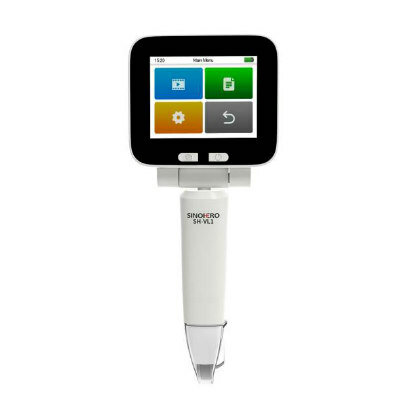
Video Laryngoscope
SH-VL1