


Understanding Post-Operative Loss of Vision
|
By HospiMedica International staff writers Posted on 28 Oct 2008 |


|
A new review of the studies, findings, and current literature regarding loss of vision after non-ocular surgery provides recommendations for surgical teams in preventing this tragic complication.
The review, written by researchers the University of Illinois (Chicago, IL, USA), covers the results of two large retrospective studies, which show that post-operative visual loss (POVL) can be as high as 4.5% in cardiac surgery and 0.2% in spine surgery. There are several causes of POVL after non-ocular surgery, including ischemic optic neuropathy, central retinal artery or vein occlusion, and cortical blindness. In fact, any interruption of blood supply to the eyes during surgery--whether caused by pressure to the eye itself or to the blood supply--or an interruption to the patient's blood flow autoregulation system can lead to POVL. One study showed that there was a higher increase of intraocular pressure (IOP) in patients whose heads were supported by pillows versus patients whose heads were supported by pins in the prone position. Another study demonstrated that as time spent in the prone position increased, IOP continued to increase as well. Some authors have suggested that IOP may increase as a result of large amounts of intravenous (IV) fluids given during surgery.
A variety of factors have been identified that contribute to the various causes of POVL, some intrinsic to the patient that can be thought of as predisposing factors, while others are related more directly to surgery and anesthesia. One study concluded that consideration should be given to establishing a minimum systolic blood pressure for each patient as well as staging long procedures and protecting eye position during surgery. Other recommendations include careful preoperative history to identify any preexisting patient risk factors that predispose to POVL; considered use of deliberate hypotension in patients at risk; avoidance of compression of the eyes, abdomen, or chest in patients placed prone; and correction of anemia and/or hypotension as early as is feasible upon identification of these risk factors. The study was published in the October 2008 issue of Neuro-Ophthalmology.
"Determining the actual overall incidence of POVL is difficult since it is not known what percentage of cases is reported,” said lead author, Molly Gilbert, M.D., an assistant professor of ophthalmology at the University of Illinois Eye and Ear Infirmary. "A wide variety of surgical interventions have been associated with POVL, including cardiopulmonary bypass, lumbar spine surgery, neck dissection, abdominal procedures, hip surgery, cholecystectomy, parathyroidectomy, prostate surgery, pleurodesis, and rotator cuff surgery.”
Related Links:
University of Illinois




The review, written by researchers the University of Illinois (Chicago, IL, USA), covers the results of two large retrospective studies, which show that post-operative visual loss (POVL) can be as high as 4.5% in cardiac surgery and 0.2% in spine surgery. There are several causes of POVL after non-ocular surgery, including ischemic optic neuropathy, central retinal artery or vein occlusion, and cortical blindness. In fact, any interruption of blood supply to the eyes during surgery--whether caused by pressure to the eye itself or to the blood supply--or an interruption to the patient's blood flow autoregulation system can lead to POVL. One study showed that there was a higher increase of intraocular pressure (IOP) in patients whose heads were supported by pillows versus patients whose heads were supported by pins in the prone position. Another study demonstrated that as time spent in the prone position increased, IOP continued to increase as well. Some authors have suggested that IOP may increase as a result of large amounts of intravenous (IV) fluids given during surgery.
A variety of factors have been identified that contribute to the various causes of POVL, some intrinsic to the patient that can be thought of as predisposing factors, while others are related more directly to surgery and anesthesia. One study concluded that consideration should be given to establishing a minimum systolic blood pressure for each patient as well as staging long procedures and protecting eye position during surgery. Other recommendations include careful preoperative history to identify any preexisting patient risk factors that predispose to POVL; considered use of deliberate hypotension in patients at risk; avoidance of compression of the eyes, abdomen, or chest in patients placed prone; and correction of anemia and/or hypotension as early as is feasible upon identification of these risk factors. The study was published in the October 2008 issue of Neuro-Ophthalmology.
"Determining the actual overall incidence of POVL is difficult since it is not known what percentage of cases is reported,” said lead author, Molly Gilbert, M.D., an assistant professor of ophthalmology at the University of Illinois Eye and Ear Infirmary. "A wide variety of surgical interventions have been associated with POVL, including cardiopulmonary bypass, lumbar spine surgery, neck dissection, abdominal procedures, hip surgery, cholecystectomy, parathyroidectomy, prostate surgery, pleurodesis, and rotator cuff surgery.”
Related Links:
University of Illinois

Gold Member
12-Channel ECG
CM1200B
Digital Radiography System (Ceiling Free)
Digix CF Series
Glucose Meter
StatStrip®
Latest Surgical Techniques News
- Needle-Free Intrauterine Anesthesia Technique Reduces Pain During IUD Insertion
- Robotic Platform Advances Fetoscopic Treatment of Twin-to-Twin Transfusion Syndrome
- Patient-Specific AI Tool Aligns X-Rays With 3D Scans For Surgical Navigation
- FDA Clears Integrated Platform for Real-Time Cardiac Imaging and Catheter Guidance
- Hydrogel Dermal Supports Healing of Complex Wounds After Cancer Surgery
- New Microcatheter Enhances Lesion Crossing in Complex Coronary Interventions
- Integrated PET/MRI and EEG Improves Presurgical Lesion Localization in Pediatric Focal Epilepsy
- Integrated Perioperative Care Model Improves Recovery in Older Surgical Patients
- New Transvaginal Transducer Improves Imaging for Gynecology and Early Obstetrics
- FDA Approves Abbott Dual-Energy Catheter for Complex AFib Cases
- Mobile 3-in-1 Imaging System Advances Intraoperative Navigation
- First Thyroid Cancer-Specific Anxiety Scale Supports Patient-Centered Care
- Tissue-Mimicking Scaffold Could Improve Rotator Cuff Repair
- Augmented Reality System Integrates Intraoperative Data for Hip Arthroscopy
- Nanobone Material Activates Natural Repair Signals to Regrow Bone
- First Robotic Dual Liver-Kidney Transplant Expands Minimally Invasive Transplant Surgery
Channels
Artificial Intelligence
view channel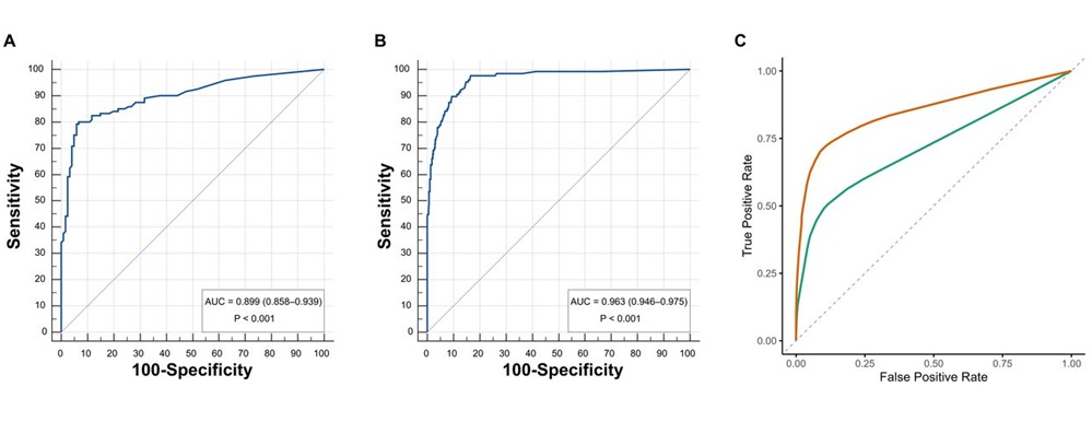
AI Improves Non-Contrast CT Interpretation for Time-Sensitive Stroke Assessment
Acute ischemic stroke occurs when a blood vessel in the brain becomes blocked, requiring rapid diagnosis to enable timely reperfusion therapy. Emergency departments often use computed tomography angiography... Read more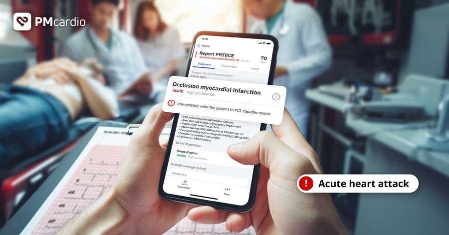
AI-Powered ECG Model Gains FDA Authorization for Emergency Cardiac Assessment
Powerful Medical’s PMcardio Queen of Hearts (ACS AI ECG Model) has received U.S. Food and Drug Administration (FDA) De Novo authorization, announced on September 15, 2026. The model is designed to identify... Read more

Critical Care
view channel
Portable Artificial Lung Technology Could Reshape Care for Chronic Lung Disease
Chronic lung disease can leave patients dependent on prolonged mechanical ventilation or awaiting transplantation, both of which carry substantial morbidity and healthcare costs. Ventilatory support may... Read more
Multimodal AI Improves Treatment Response Prediction in Lung Cancer
Immunotherapy has transformed care for advanced non-small cell lung cancer (NSCLC), yet only a minority of patients achieve durable benefit. Clinicians still struggle to predict who will respond, and treatment... Read more
New Approach Helps Identify Materials for More Reliable Wearable Electronics
Wearable health monitors and electronic skin must maintain stable performance while being stretched and bent. Molecular single crystals are promising for such stretchable electronics, yet the microscale... Read more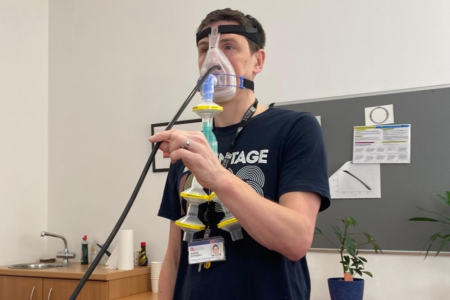
Scotland Adopts Respiratory Infection Control Guidance Informed by Aerosol Research
Preventing respiratory infection transmission in hospitals remains a core patient and staff safety priority. Many precautions have hinged on the assumption that certain clinical interventions create high-risk... Read morePatient Care
view channel
EHR Model Flags High-Risk Periods in Patients with Metastatic Breast Cancer
Determining when patients with metastatic breast cancer are nearing the end of life remains difficult, often leading to intensive interventions with limited benefit. Prognostic uncertainty can delay goals-of-care... Read more
Virtual Reality Helps Claustrophobic Patients Complete Cardiac PET/CT Scans
Anxiety and claustrophobia—intense fear of confined spaces—often prevent patients from completing cardiac positron emission tomography/computed tomography (PET/CT). Aborted scans delay diagnosis, consume... Read more

Medical Imaging
view channel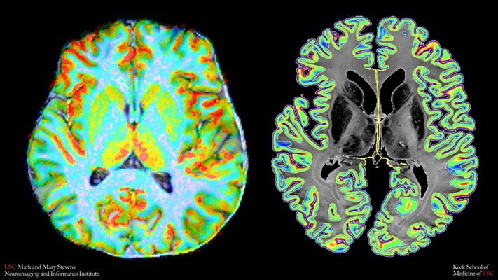
New MRI Index Reveals How Brain Blood Flow Supports Cellular Energy Needs
The human brain has minimal energy reserves and relies on tightly regulated blood flow to meet its high metabolic demands. However, clinicians have lacked noninvasive tools to assess how cortical perfusion... Read more
Structure-Aware AI Aims to Improve Reliability of Medical Image Analysis
Clinicians increasingly rely on medical imaging to guide diagnosis and treatment, but machine learning systems used to analyze these images can be unreliable and difficult to interpret. Small, heterogeneous... Read more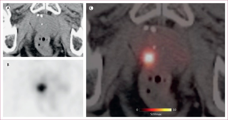

Health IT
view channel
New Tablet Platform Extends Stroke Neurorehabilitation Beyond the Clinic
Continuity and intensity of neurorehabilitation after stroke are critical for functional recovery, but access to ongoing therapy at home often remains limited. Hospitals and outpatient programs therefore... Read more
Smartphone App Improves Blood Pressure Control in Hypertension
Hypertension, or high blood pressure, is a major modifiable driver of cardiovascular, renal, and cerebrovascular morbidity. Despite effective therapies, many adults fail to achieve control, leading to... Read more

Point of Care
view channel
Continuous Glucose Monitoring Identifies Cardiometabolic Risk in Adults Without Diabetes
Dysglycemia—abnormal blood glucose regulation—can fluctuate throughout the day and often escape conventional screening. Clinicians typically rely on fasting plasma glucose and hemoglobin A1c, which offer... Read more
Microneedle Patch Enables At-Home Monitoring of Acute Kidney Injury
Kidney disease is often silent in its early stages, making timely detection difficult outside clinical settings. Early biomarkers such as neutrophil gelatinase–associated lipocalin can require blood draws... Read more
Business
view channel
Medtronic Receives FDA Clearance for Vessel-Sealing Instrument for Robotic Surgery
As robotic-assisted surgery expands across U.S. hospitals, teams increasingly seek energy instruments with the familiarity and performance of tools used in open and laparoscopic procedures.... Read more
Robotic Single-Port System Gains CE Mark for Transvaginal Gynecologic Procedures
Intuitive (Sunnyvale, CA, USA) announced that it has received CE mark approval for use of the da Vinci SP Single Port surgical system in transvaginal gynecologic procedures, marking the first such indication... Read more