


3D Skin Printer Covers Large Burn Wound Areas 
|
By HospiMedica International staff writers Posted on 20 Feb 2020 |
|
")
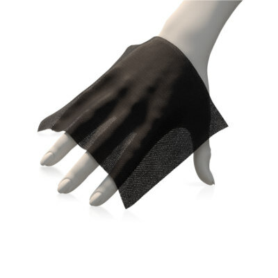
Image: The disposable deposition head of the bio-ink printer (Photo courtesy of UT)
A new study describes how a novel bio-ink printer forms a uniform sheet of mesenchymal stroma cells (MSCs) over burn areas, promoting skin regeneration and reducing scarring.
Under development at the University of Toronto (UT; Canada) and Sunnybrook Research Institute (Sunnybrook; Toronto, Canada), the current prototype of the hand-held device includes a microfluidic print-head that forms tissue in situ, depositing and setting the MSCs in place in two minutes or less. A soft wheel follows the track of the print-head, allowing better control for wider wounds. To ensure sterilization, the print-head is disposable.
In porcine pre-clinical models of a full-thickness burn, the researchers successfully delivered MSCs and stromal cell-containing fibrin sheets directly to the wound bed, improving re-epithelialization, dermal cell repopulation, and neovascularization. The researchers also demonstrated conformal delivery to surfaces that were inclined up to 45°, indicating that the 3D printer could be introduced in a clinical setting to improve dermal and epidermal regeneration. The study was published on February 4, 2020, in Biofabrication.
“Full-thickness burns are characterized by the destruction of both the outermost and innermost layers of the skin; these burns often cover a significant portion of the body. With big burns, you don’t have sufficient healthy skin available,” said co-senior author Marc Jeschke, MD, director of the Sunnybrook Ross Tilley Burn Centre. “Once it’s used in an operating room, I think this printer will be a game changer in saving lives. With a device like this, it could change the entirety of how we practice burn and trauma care.”
A full-thickness burn (also known as third- and fourth degree burns) occurs when both the epidermis and dermis are destroyed and the burn extends down into the subcutaneous tissue, including fat, muscles and even bones. For full-thickness burns, generally the skin will either be white, black, brown, charred, or leathery in appearance. Often eschar--dry, black, necrotic tissue--will form around the wound. Since nerve endings are destroyed along with the dermis, these wounds are typically painless. Another contributing factor to burn severity is its extension.
Related Links:
University of Toronto
Sunnybrook Research Institute
Gold Member
12-Channel ECG
CM1200B
Gold Member
SARS‑CoV‑2/Flu A/Flu B/RSV Sample-To-Answer Test
SARS‑CoV‑2/Flu A/Flu B/RSV Cartridge (CE-IVD)
New
Antimicrobial Barrier Dressing
ACTICOAT FLEX
New
Bioengineered Collagen Implant
Tapestry Biointegrative Implant
Latest Surgical Techniques News
- Easy-To-Apply Gel Could Prevent Formation of Post-Surgical Abdominal Adhesions
- Groundbreaking Leadless Pacemaker to Prevent Invasive Surgeries for Children
- Spectroscopy Technique Improves Surgery for Pediatric Epilepsy Patients
- Bioengineered Arteries Show Promise for Cardiovascular Surgery
- Online Tool Guides Surgical Decisions for Gallbladder Cancer
- Innovative Technology Enables Rapid Life-Saving Surgical Leak Detection
- First-Of-Its-Kind Bioresorbable Implant to Help Children with Rare Respiratory Disease
- Screw-Shaped Magnetic Microrobots to Transform Treatment for Patients with Inoperable Blood Clots
- "Ultra-Rapid" Testing in the OR Could Enable Accurate Removal of Brain Tumors
- Automated Endoscopic Device Obtains Improved Biopsy Results in Single Pass
- World's First Machine Learning Model Combats Wrong-Site Surgery
- Novel Method Combining Heart Biopsy and Device Implantation Reduces Complications Risk
- New Surface Coating Could Prevent Blood Clotting in Medical Devices and Implants
- Dumbbell-Shaped Thrombectomy Device Offers Novel Approach to Cerebral Venous Sinus Thrombosis Treatment
- Novel Catheter Mimics Snake Teeth to Grab Blood Clots
- New Laparoscopic Imaging Technique Accurately Maps Biological Tissue for Minimally Invasive Surgery
Channels
Artificial Intelligence
view channel")
Innovative Risk Score Predicts Heart Attack or Stroke in Kidney Transplant Candidates
Heart researchers have utilized an innovative risk assessment score to accurately predict whether patients being evaluated for kidney transplants are at risk for future major cardiac events, such as a... Read more")
AI Algorithm Detects Early-Stage Metabolic-Associated Steatotic Liver Disease Using EHRs
Liver disease, which is treatable when detected early, often goes unnoticed until it reaches advanced stages. Metabolic-associated steatotic liver disease (MASLD), the most prevalent form of liver disease,... Read moreCritical Care
view channel")
Novel Coating Significantly Extends Longevity of Implantable Biosensors
Wearable and implantable biosensors capable of accurately detecting biological molecules in a non-invasive or minimally invasive way offer enormous potential for monitoring patients’ health and their responses... Read more")
Nanogel-Based Drug Delivery Technology to Improve UTI Treatment
Urinary tract infections (UTIs) are not only widespread and costly but also highly debilitating, significantly impacting the quality of life for those affected. The antibiotics commonly used to treat UTIs... Read more")
New IV Pole Improves Safety and Ease of Administering IV Medications at Hospital Bedside
Preventable medication errors affect around 500,000 hospitalized patients in the U.S. every year. A significant portion of these errors occur with intravenous (IV) smart pumps, which require a precise... Read more")
Patient Care
view channel")
Portable Biosensor Platform to Reduce Hospital-Acquired Infections
Approximately 4 million patients in the European Union acquire healthcare-associated infections (HAIs) or nosocomial infections each year, with around 37,000 deaths directly resulting from these infections,... Read moreFirst-Of-Its-Kind Portable Germicidal Light Technology Disinfects High-Touch Clinical Surfaces in Seconds
Reducing healthcare-acquired infections (HAIs) remains a pressing issue within global healthcare systems. In the United States alone, 1.7 million patients contract HAIs annually, leading to approximately... Read more")
Surgical Capacity Optimization Solution Helps Hospitals Boost OR Utilization
An innovative solution has the capability to transform surgical capacity utilization by targeting the root cause of surgical block time inefficiencies. Fujitsu Limited’s (Tokyo, Japan) Surgical Capacity... Read more")
Game-Changing Innovation in Surgical Instrument Sterilization Significantly Improves OR Throughput
A groundbreaking innovation enables hospitals to significantly improve instrument processing time and throughput in operating rooms (ORs) and sterile processing departments. Turbett Surgical, Inc.... Read moreHealth IT
view channel")
Printable Molecule-Selective Nanoparticles Enable Mass Production of Wearable Biosensors
The future of medicine is likely to focus on the personalization of healthcare—understanding exactly what an individual requires and delivering the appropriate combination of nutrients, metabolites, and... Read more")
Smartwatches Could Detect Congestive Heart Failure
Diagnosing congestive heart failure (CHF) typically requires expensive and time-consuming imaging techniques like echocardiography, also known as cardiac ultrasound. Previously, detecting CHF by analyzing... Read more")
")
Point of Care
view channel")
Handheld, Sound-Based Diagnostic System Delivers Bedside Blood Test Results in An Hour
Patients who go to a doctor for a blood test often have to contend with a needle and syringe, followed by a long wait—sometimes hours or even days—for lab results. Scientists have been working hard to... Read more")
Smartphone-Enabled, Paper-Based Quantitative Diagnostic Platform Transforms POC Testing
Point-of-care diagnostics are crucial for public health, offering rapid, on-site testing that enables prompt diagnosis and treatment. This is especially valuable in remote or underserved regions where... Read moreBusiness
view channel")
Becton Dickinson to Spin Out Biosciences and Diagnostic Solutions Business
Becton, Dickinson and Company (BD, Franklin Lakes, NJ, USA), has announced that its board of directors has unanimously authorized BD management to pursue a plan to separate BD's Biosciences and Diagnostic... Read more")
")
")