Two-Step Approach Helps Repair Herniated Discs 
|
By HospiMedica International staff writers Posted on 01 Apr 2020 |
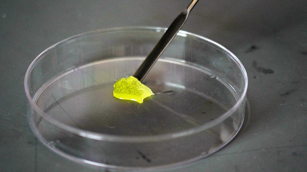
Image: This riboflavin infused collagen gel stiffens into a solid when illuminated (Photo courtesy of Cornell University)
A combined therapeutic approach helps heal annulus fibrosus defects, restore nucleus pulposus hydration, and maintain native torsional and compressive stiffness, according to a new study.
Developed by researchers at Cornell University (Cornell; Ithaca, NY, USA), the Hospital for Special Surgery (HSS, New York, NY, USA), and other institutions, the acellular, tissue-engineered technique is designed to prevent degeneration after a discectomy procedure. The two-step technique involves first injecting hyaluronic acid into the inner region of the disc--the nucleus pulposus--and subsequently applying a photo-crosslinked collagen patch to the healed outer annulus fibrosus defects. The collagen patch also incorporates riboflavin, a photoactive vitamin B derivative.
Instead of suturing the ruptured disc, the riboflavin is activated by shining a light on it, which causes the thick gel to stiffen into a solid. Importantly, the gel provides a fertile ground for cells to grow new tissue, sealing the defect better than any suture could. The researchers found the technique successfully healed the damage to the annulus fibrosus, restored disc height, and maintained mechanical performance and biomechanical support of the spine up to six weeks after injury. The study was published on March 11, 2020, in Science Translational Medicine.
“This is really a new avenue and a whole new approach to treating people who have herniated discs, other than walking around with a big hole in their intervertebral disc and hoping that it doesn’t re-herniate or continue to degenerate,” said senior author Lawrence Bonassar, PhD, of Cornell University. “The idea is, if you have a herniation and you’ve lost some material from the nucleus, now we can re-inflate the disc with this hyaluronic acid gel and put the collagen cross-linking seal on the outside. Now we’ve refilled the tire and sealed it.”
The intervertebral discs are composed of two parts: a stiffer external tissue called the annulus fibrosis, and a softer, gelatinous material in the center, the nucleus pulposus, which keeps the disc pressurized and able to hold its shape and height during physical movement. If the outer layer ruptures, the jelly-like nucleus pulposus leaks out, causing inflammation in the nerve root or the spinal cord itself.
Related Links:
Cornell University
Hospital for Special Surgery
Latest Surgical Techniques News
- Bioprinted Aortas Offer New Hope for Vascular Repair
- Early TAVR Intervention Reduces Cardiovascular Events in Asymptomatic Aortic Stenosis Patients
- New Procedure Found Safe and Effective for Patients Undergoing Transcatheter Mitral Valve Replacement
- No-Touch Vein Harvesting Reduces Graft Failure Risk for Heart Bypass Patients
- DNA Origami Improves Imaging of Dense Pancreatic Tissue for Cancer Detection and Treatment
- Pioneering Sutureless Coronary Bypass Technology to Eliminate Open-Chest Procedures
- Intravascular Imaging for Guiding Stent Implantation Ensures Safer Stenting Procedures
- World's First AI Surgical Guidance Platform Allows Surgeons to Measure Success in Real-Time
- AI-Generated Synthetic Scarred Hearts Aid Atrial Fibrillation Treatment
- New Class of Bioadhesives to Connect Human Tissues to Long-Term Medical Implants
- New Transcatheter Valve Found Safe and Effective for Treating Aortic Regurgitation
- Minimally Invasive Valve Repair Reduces Hospitalizations in Severe Tricuspid Regurgitation Patients
- Tiny Robotic Tools Powered by Magnetic Fields to Enable Minimally Invasive Brain Surgery
- Magnetic Tweezers Make Robotic Surgery Safer and More Precise
- AI-Powered Surgical Planning Tool Improves Pre-Op Planning
- Novel Sensing System Restores Missing Sense of Touch in Minimally Invasive Surgery
Channels
Critical Care
view channel
Mechanosensing-Based Approach Offers Promising Strategy to Treat Cardiovascular Fibrosis
Cardiac fibrosis, which involves the stiffening and scarring of heart tissue, is a fundamental feature of nearly every type of heart disease, from acute ischemic injuries to genetic cardiomyopathies.... Read more
AI Interpretability Tool for Photographed ECG Images Offers Pixel-Level Precision
The electrocardiogram (ECG) is a crucial diagnostic tool in modern medicine, used to detect heart conditions such as arrhythmias and structural abnormalities. Every year, millions of ECGs are performed... Read morePatient Care
view channel
Portable Biosensor Platform to Reduce Hospital-Acquired Infections
Approximately 4 million patients in the European Union acquire healthcare-associated infections (HAIs) or nosocomial infections each year, with around 37,000 deaths directly resulting from these infections,... Read moreFirst-Of-Its-Kind Portable Germicidal Light Technology Disinfects High-Touch Clinical Surfaces in Seconds
Reducing healthcare-acquired infections (HAIs) remains a pressing issue within global healthcare systems. In the United States alone, 1.7 million patients contract HAIs annually, leading to approximately... Read more
Surgical Capacity Optimization Solution Helps Hospitals Boost OR Utilization
An innovative solution has the capability to transform surgical capacity utilization by targeting the root cause of surgical block time inefficiencies. Fujitsu Limited’s (Tokyo, Japan) Surgical Capacity... Read more
Game-Changing Innovation in Surgical Instrument Sterilization Significantly Improves OR Throughput
A groundbreaking innovation enables hospitals to significantly improve instrument processing time and throughput in operating rooms (ORs) and sterile processing departments. Turbett Surgical, Inc.... Read moreHealth IT
view channel
Printable Molecule-Selective Nanoparticles Enable Mass Production of Wearable Biosensors
The future of medicine is likely to focus on the personalization of healthcare—understanding exactly what an individual requires and delivering the appropriate combination of nutrients, metabolites, and... Read more
Smartwatches Could Detect Congestive Heart Failure
Diagnosing congestive heart failure (CHF) typically requires expensive and time-consuming imaging techniques like echocardiography, also known as cardiac ultrasound. Previously, detecting CHF by analyzing... Read moreBusiness
view channel
Expanded Collaboration to Transform OR Technology Through AI and Automation
The expansion of an existing collaboration between three leading companies aims to develop artificial intelligence (AI)-driven solutions for smart operating rooms with sophisticated monitoring and automation.... Read more


