Delayed Fluorescence Imaging Method Could Enable Effective Surgical Tumor Removal
|
By HospiMedica International staff writers Posted on 12 Oct 2022 |

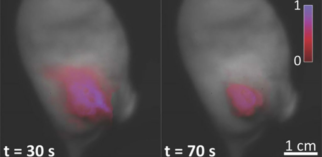
In the surgical treatment of cancer, distinguishing between tumors and healthy tissues is critical. Fluorescent markers can help to do this, by enhancing the contrast of tumors during surgery. Some markers show a phenomenon called “delayed fluorescence” (DF) which relies on detecting “hypoxia” (or low oxygen concentration), a condition often presented by tumors. The real-time imaging of hypoxia can provide a high contrast between tumors and healthy cells. This can allow surgeons to remove the tumor effectively. However, real-time hypoxia imaging for surgical guidance has not yet been achieved. In a new study, researchers have proposed an optical imaging system that enables real-time imaging of tissue oxygen concentration for tumors presenting chronic or transient hypoxia. The team achieved this by making use of an endogenous molecule called protoporphyrin IX (PpIX), which exhibits DF in the red to near-infrared region.
The technical challenge in detecting DF is due to its low intensity; background noise makes it difficult to detect without a single photon detector. The team of researchers at Dartmouth College (Hanover, NH, USA) overcame this problem using a highly sensitive time-gated imaging system, which allows signal detection within a specified time window only. This greatly reduces the background noise and enables a wide-field direct mapping of oxygen partial pressure (pO2) changes with the acquired DF signal. The result is real-time metabolic information, a useful map for surgical guidance.
The team demonstrated the efficacy of the technique using mice models of pancreatic cancer, which exhibited hypoxic tumors. The DF signal obtained from the cancerous cells was over five times stronger than that from surrounding healthy oxygenated tissues. The signal contrast was further enhanced when the tissues were palpated before imaging to further enhance transient hypoxia. The imaging of pO2 in tissues could also enable control of tissue metabolism. This, in turn, would help us better understand the biochemistry involved in oxygen supply and consumption.
“This is a truly unique reporter of the local oxygen partial pressure in tissues. PpIX is endogenously synthesized by mitochondria in most tissues, and the particular property of DF emission is directly related to low microenvironmental oxygen concentration,” explained Brian Pogue, Chair of Medical Physics at University of Wisconsin-Madison, Adjunct Professor of Engineering Sciences at Dartmouth College, and senior author of the study. “Healthy cells will show little to no DF, because it is quenched in the presence of molecular oxygen.”
“Acquiring both prompt and delayed fluorescence in a rapid sequential cycle allowed for imaging oxygen levels in a way that was independent of the PpIX concentration,” said Lead author Arthur Petusseau, a doctoral candidate in Engineering Sciences at Dartmouth College.
“The results reported by Petusseau’s team suggest hypoxia imaging as an efficient approach to identifying tumors in cancer treatment,” said Frédéric Leblond, Professor of Engineering Physics at Polytechnique Montréal and JBO Associate Editor. “PpIX DF detection uses a known clinical dye and an already-approved in-human marker, with great potential for surgical guidance, and more.”
Related Links:
Dartmouth College
Latest Surgical Techniques News
- Bioprinted Aortas Offer New Hope for Vascular Repair
- Early TAVR Intervention Reduces Cardiovascular Events in Asymptomatic Aortic Stenosis Patients
- New Procedure Found Safe and Effective for Patients Undergoing Transcatheter Mitral Valve Replacement
- No-Touch Vein Harvesting Reduces Graft Failure Risk for Heart Bypass Patients
- DNA Origami Improves Imaging of Dense Pancreatic Tissue for Cancer Detection and Treatment
- Pioneering Sutureless Coronary Bypass Technology to Eliminate Open-Chest Procedures
- Intravascular Imaging for Guiding Stent Implantation Ensures Safer Stenting Procedures
- World's First AI Surgical Guidance Platform Allows Surgeons to Measure Success in Real-Time
- AI-Generated Synthetic Scarred Hearts Aid Atrial Fibrillation Treatment
- New Class of Bioadhesives to Connect Human Tissues to Long-Term Medical Implants
- New Transcatheter Valve Found Safe and Effective for Treating Aortic Regurgitation
- Minimally Invasive Valve Repair Reduces Hospitalizations in Severe Tricuspid Regurgitation Patients
- Tiny Robotic Tools Powered by Magnetic Fields to Enable Minimally Invasive Brain Surgery
- Magnetic Tweezers Make Robotic Surgery Safer and More Precise
- AI-Powered Surgical Planning Tool Improves Pre-Op Planning
- Novel Sensing System Restores Missing Sense of Touch in Minimally Invasive Surgery
Channels
Critical Care
view channel
Mechanosensing-Based Approach Offers Promising Strategy to Treat Cardiovascular Fibrosis
Cardiac fibrosis, which involves the stiffening and scarring of heart tissue, is a fundamental feature of nearly every type of heart disease, from acute ischemic injuries to genetic cardiomyopathies.... Read more
AI Interpretability Tool for Photographed ECG Images Offers Pixel-Level Precision
The electrocardiogram (ECG) is a crucial diagnostic tool in modern medicine, used to detect heart conditions such as arrhythmias and structural abnormalities. Every year, millions of ECGs are performed... Read morePatient Care
view channel
Portable Biosensor Platform to Reduce Hospital-Acquired Infections
Approximately 4 million patients in the European Union acquire healthcare-associated infections (HAIs) or nosocomial infections each year, with around 37,000 deaths directly resulting from these infections,... Read moreFirst-Of-Its-Kind Portable Germicidal Light Technology Disinfects High-Touch Clinical Surfaces in Seconds
Reducing healthcare-acquired infections (HAIs) remains a pressing issue within global healthcare systems. In the United States alone, 1.7 million patients contract HAIs annually, leading to approximately... Read more
Surgical Capacity Optimization Solution Helps Hospitals Boost OR Utilization
An innovative solution has the capability to transform surgical capacity utilization by targeting the root cause of surgical block time inefficiencies. Fujitsu Limited’s (Tokyo, Japan) Surgical Capacity... Read more
Game-Changing Innovation in Surgical Instrument Sterilization Significantly Improves OR Throughput
A groundbreaking innovation enables hospitals to significantly improve instrument processing time and throughput in operating rooms (ORs) and sterile processing departments. Turbett Surgical, Inc.... Read moreHealth IT
view channel
Printable Molecule-Selective Nanoparticles Enable Mass Production of Wearable Biosensors
The future of medicine is likely to focus on the personalization of healthcare—understanding exactly what an individual requires and delivering the appropriate combination of nutrients, metabolites, and... Read more
Smartwatches Could Detect Congestive Heart Failure
Diagnosing congestive heart failure (CHF) typically requires expensive and time-consuming imaging techniques like echocardiography, also known as cardiac ultrasound. Previously, detecting CHF by analyzing... Read moreBusiness
view channel
Expanded Collaboration to Transform OR Technology Through AI and Automation
The expansion of an existing collaboration between three leading companies aims to develop artificial intelligence (AI)-driven solutions for smart operating rooms with sophisticated monitoring and automation.... Read more


