Narrow-Band Imaging Detects Lesions Overlooked by White-Light Imaging during Endoscopic Examinations
|
By HospiMedica International staff writers Posted on 22 Dec 2023 |
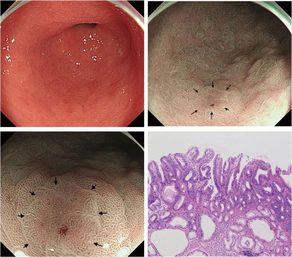
Helicobacter pylori infection is a well-recognized significant risk factor for developing gastric cancer, making regular screenings for early detection particularly vital in high-prevalence areas. While modern endoscopy techniques allow for the removal of significant neoplasms, the focus is increasingly on the early identification of smaller lesions. Despite these advancements, the detection of subtle epithelial neoplasms, which represent about a quarter of all newly diagnosed small neoplasms, poses a challenge. Now, new research suggests that employing magnifying narrow-band imaging at low magnification (LM-NBI) following routine white-light imaging (WLI) could improve the detection rate of lesions that WLI might miss.
In order to address a research gap regarding the effectiveness of periodic LM-NBI screenings compared to conventional magnifying endoscopy (CE), a study was undertaken at Nagashima Clinic (Yamagata, Japan). This study aimed to ascertain the effectiveness of annual endoscopic exams utilizing LM-NBI for the entire stomach after initial WLI, focusing on identifying gastric neoplasms. In both the LM-NBI and CE groups, the initial endoscopy standardized the baseline, followed by up to five annual endoscopies. The inaugural annual examination for the LM-NBI group occurred between April 2019 and March 2020, and for the CE group, it took place between April 2015 and March 2016. A retrospective examination of medical records was conducted to evaluate gastric tumor detection.
The study's findings showed that among the 388 patients in the LM-NBI group and the 381 in the CE group, 15 and 5 patients, respectively, were diagnosed with gastric neoplasia, excluding one case of mucosa-associated lymphoid tissue lymphoma. Each detected case was classified as epithelial neoplasia. The endoscopic procedures were safely executed without any complications necessitating further intervention. According to the Cox proportional hazards model, there was a hazard ratio of 2.78 (95% CI, 1.01–7.64). Kaplan–Meier analysis (p = 0.039, log-rank test) demonstrated that annual LM-NBI was more effective than CE in detecting gastric neoplasia.
This historical case-control study crucially demonstrates a nearly threefold increase in the detection rate of neoplastic lesions in the stomach through annual LM-NBI following WLI compared to CE. It is the first study to confirm the effectiveness of annual endoscopy using LM-NBI in spotting gastric neoplasms, especially in areas with map-like redness or in atrophic/metaplastic mucosa. The study also emphasizes the value of conducting both WLI and NBI examinations in tandem to ensure thorough lesion detection, acknowledging the tendency of observers to concentrate on color changes in WLI.
“While LM-NBI successfully identified lesions overlooked by WLI, its efficacy in detecting diffuse-type carcinomas remains uncertain, especially for signet ring cell carcinomas,” noted Ryuichi Nagashima, sole author of the study. “Further studies are needed to clarify the superiority of LM-NBI over other image-enhanced endoscopies, such as blue laser imaging bright and linked color imaging, to establish its optimal role in clinical practice.”
Related Links:
Nagashima Clinic
Latest Surgical Techniques News
- Pioneering Sutureless Coronary Bypass Technology to Eliminate Open-Chest Procedures
- Intravascular Imaging for Guiding Stent Implantation Ensures Safer Stenting Procedures
- World's First AI Surgical Guidance Platform Allows Surgeons to Measure Success in Real-Time
- AI-Generated Synthetic Scarred Hearts Aid Atrial Fibrillation Treatment
- New Class of Bioadhesives to Connect Human Tissues to Long-Term Medical Implants
- New Transcatheter Valve Found Safe and Effective for Treating Aortic Regurgitation
- Minimally Invasive Valve Repair Reduces Hospitalizations in Severe Tricuspid Regurgitation Patients
- Tiny Robotic Tools Powered by Magnetic Fields to Enable Minimally Invasive Brain Surgery
- Magnetic Tweezers Make Robotic Surgery Safer and More Precise
- AI-Powered Surgical Planning Tool Improves Pre-Op Planning
- Novel Sensing System Restores Missing Sense of Touch in Minimally Invasive Surgery
- Headset-Based AR Navigation System Improves EVD Placement
- Higher Electrode Density Improves Epilepsy Surgery by Pinpointing Where Seizures Begin
- Open-Source Tool Optimizes Placement of Visual Brain Implants
- Easy-To-Apply Gel Could Prevent Formation of Post-Surgical Abdominal Adhesions
- Groundbreaking Leadless Pacemaker to Prevent Invasive Surgeries for Children
Channels
Critical Care
view channel
Ingestible Smart Capsule for Chemical Sensing in the Gut Moves Closer to Market
Intestinal gases are associated with several health conditions, including colon cancer, irritable bowel syndrome, and inflammatory bowel disease, and they have the potential to serve as crucial biomarkers... Read moreNovel Cannula Delivery System Enables Targeted Delivery of Imaging Agents and Drugs
Multiphoton microscopy has become an invaluable tool in neuroscience, allowing researchers to observe brain activity in real time with high-resolution imaging. A crucial aspect of many multiphoton microscopy... Read more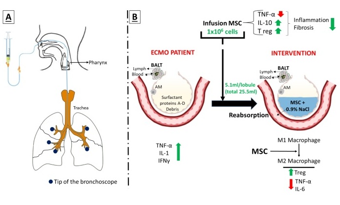
Novel Intrabronchial Method Delivers Cell Therapies in Critically Ill Patients on External Lung Support
Until now, administering cell therapies to patients on extracorporeal membrane oxygenation (ECMO)—a life-support system typically used for severe lung failure—has been nearly impossible.... Read morePatient Care
view channel
Portable Biosensor Platform to Reduce Hospital-Acquired Infections
Approximately 4 million patients in the European Union acquire healthcare-associated infections (HAIs) or nosocomial infections each year, with around 37,000 deaths directly resulting from these infections,... Read moreFirst-Of-Its-Kind Portable Germicidal Light Technology Disinfects High-Touch Clinical Surfaces in Seconds
Reducing healthcare-acquired infections (HAIs) remains a pressing issue within global healthcare systems. In the United States alone, 1.7 million patients contract HAIs annually, leading to approximately... Read more
Surgical Capacity Optimization Solution Helps Hospitals Boost OR Utilization
An innovative solution has the capability to transform surgical capacity utilization by targeting the root cause of surgical block time inefficiencies. Fujitsu Limited’s (Tokyo, Japan) Surgical Capacity... Read more
Game-Changing Innovation in Surgical Instrument Sterilization Significantly Improves OR Throughput
A groundbreaking innovation enables hospitals to significantly improve instrument processing time and throughput in operating rooms (ORs) and sterile processing departments. Turbett Surgical, Inc.... Read moreHealth IT
view channel
Printable Molecule-Selective Nanoparticles Enable Mass Production of Wearable Biosensors
The future of medicine is likely to focus on the personalization of healthcare—understanding exactly what an individual requires and delivering the appropriate combination of nutrients, metabolites, and... Read more
Smartwatches Could Detect Congestive Heart Failure
Diagnosing congestive heart failure (CHF) typically requires expensive and time-consuming imaging techniques like echocardiography, also known as cardiac ultrasound. Previously, detecting CHF by analyzing... Read moreBusiness
view channel
Expanded Collaboration to Transform OR Technology Through AI and Automation
The expansion of an existing collaboration between three leading companies aims to develop artificial intelligence (AI)-driven solutions for smart operating rooms with sophisticated monitoring and automation.... Read more


